4UW0

Low resolution structure of WbdD with C-terminal bundle ordered to residue 505


Experimental Data Snapshot

  • Method: X-RAY DIFFRACTION
  • Resolution: 3.87 Å
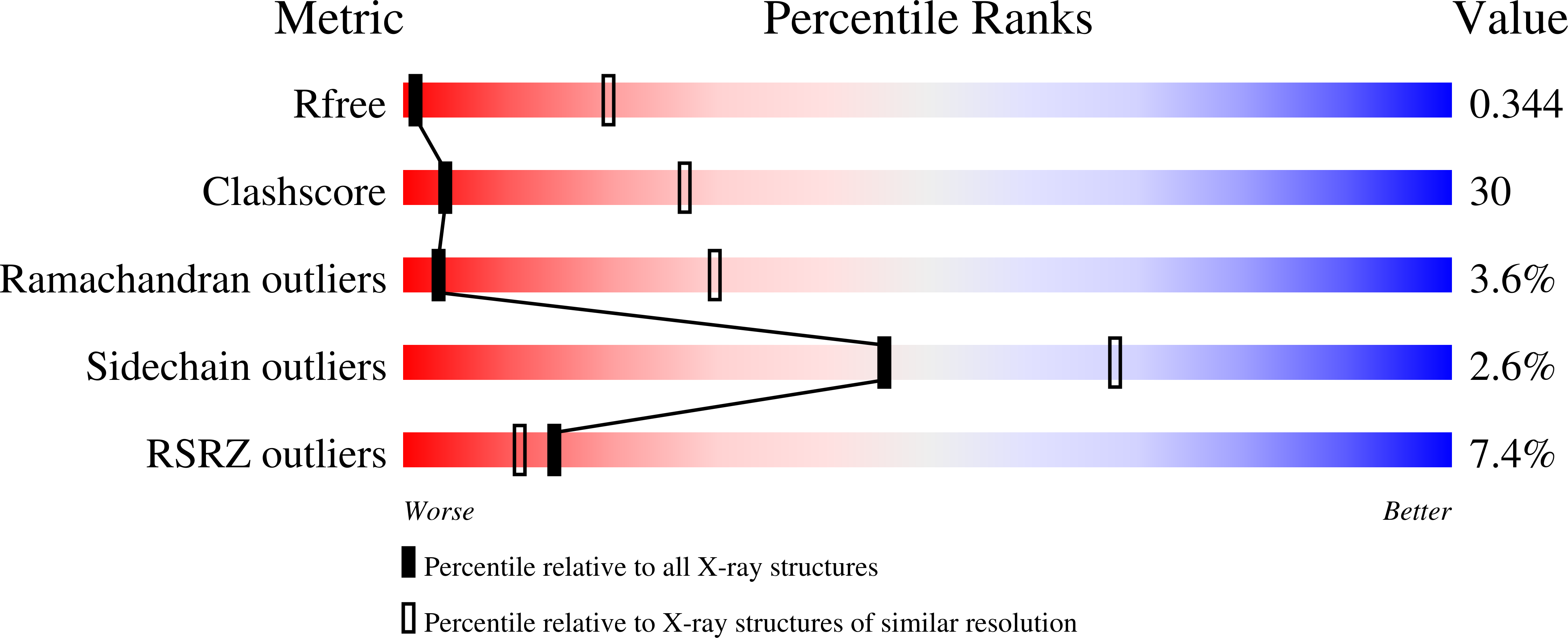
  • R-Value Free: 0.341 
  • R-Value Work: 0.268 
  • R-Value Observed: 0.271 

wwPDB Validation   3D Report Full Report


Ligand Structure Quality Assessment 


This is version 1.6 of the entry. See complete history


Literature

A Coiled-Coil Domain Acts as a Molecular Ruler to Regulate O-Antigen Chain Length in Lipopolysaccharide.

Hagelueken, G.Clarke, B.R.Huang, H.Tuukkanen, A.Danciu, I.Svergun, D.I.Hussain, R.Liu, H.Whitfield, C.Naismith, J.H.

(2015) Nat Struct Mol Biol 22: 50

  • DOI: https://doi.org/10.1038/nsmb.2935
  • Primary Citation of Related Structures:  
    4UW0

  • PubMed Abstract: 

    Long-chain bacterial polysaccharides have important roles in pathogenicity. In Escherichia coli O9a, a model for ABC transporter-dependent polysaccharide assembly, a large extracellular carbohydrate with a narrow size distribution is polymerized from monosaccharides by a complex of two proteins, WbdA (polymerase) and WbdD (terminating protein). Combining crystallography and small-angle X-ray scattering, we found that the C-terminal domain of WbdD contains an extended coiled-coil that physically separates WbdA from the catalytic domain of WbdD. The effects of insertions and deletions in the coiled-coil region were analyzed in vivo, revealing that polymer size is controlled by varying the length of the coiled-coil domain. Thus, the coiled-coil domain of WbdD functions as a molecular ruler that, along with WbdA:WbdD stoichiometry, controls the chain length of a model bacterial polysaccharide.


  • Organizational Affiliation

    Biomedical Sciences Research Complex, University of St Andrews, St Andrews, Fife, KY16 9ST, UK.


Macromolecules
Find similar proteins by:  (by identity cutoff)  |  3D Structure
Entity ID: 1
MoleculeChains Sequence LengthOrganismDetailsImage
WBDD505Escherichia coliMutation(s): 0 
Gene Names: EC3426_03072
Entity Groups  
Sequence Clusters30% Identity50% Identity70% Identity90% Identity95% Identity100% Identity
Sequence Annotations
Expand
  • Reference Sequence
Small Molecules
Ligands 1 Unique
IDChains Name / Formula / InChI Key2D Diagram3D Interactions
SAM
Query on SAM

Download Ideal Coordinates CCD File 
B [auth A]S-ADENOSYLMETHIONINE
C15 H22 N6 O5 S
MEFKEPWMEQBLKI-FCKMPRQPSA-N
Experimental Data & Validation

Experimental Data

  • Method: X-RAY DIFFRACTION
  • Resolution: 3.87 Å
  • R-Value Free: 0.341 
  • R-Value Work: 0.268 
  • R-Value Observed: 0.271 
  • Space Group: I 2 3
Unit Cell:
Length ( Å )Angle ( ˚ )
a = 181.29α = 90
b = 181.29β = 90
c = 181.29γ = 90
Software Package:
Software NamePurpose
REFMACrefinement

Structure Validation

View Full Validation Report


Ligand Structure Quality Assessment 


Entry History 

Deposition Data

Revision History  (Full details and data files)

  • Version 1.0: 2014-08-27
    Type: Initial release
  • Version 1.1: 2014-12-10
    Changes: Database references
  • Version 1.2: 2014-12-31
    Changes: Database references
  • Version 1.3: 2015-01-21
    Changes: Database references
  • Version 1.4: 2018-04-25
    Changes: Advisory, Data collection
  • Version 1.5: 2019-09-18
    Changes: Data collection, Database references, Source and taxonomy
  • Version 1.6: 2024-05-08
    Changes: Advisory, Data collection, Database references, Derived calculations, Other