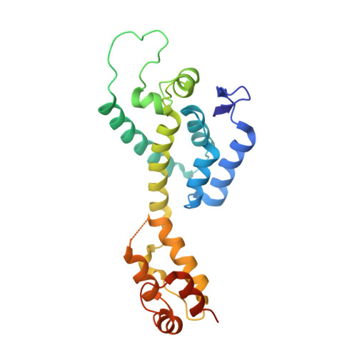
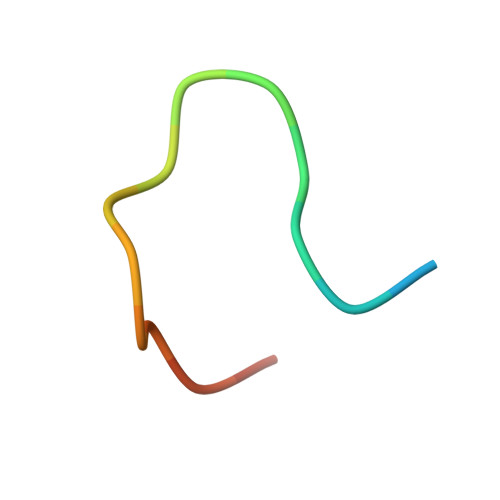
4WYM

Structural basis of HIV-1 capsid recognition by CPSF6


Experimental Data Snapshot

  • Method: X-RAY DIFFRACTION
  • Resolution: 2.60 Å
  • R-Value Free: 0.259 
  • R-Value Work: 0.219 
  • R-Value Observed: 0.219 

Starting Model: experimental
View more details

wwPDB Validation   3D Report Full Report


This is version 1.4 of the entry. See complete history


Literature

Structural basis of HIV-1 capsid recognition by PF74 and CPSF6.

Bhattacharya, A.Alam, S.L.Fricke, T.Zadrozny, K.Sedzicki, J.Taylor, A.B.Demeler, B.Pornillos, O.Ganser-Pornillos, B.K.Diaz-Griffero, F.Ivanov, D.N.Yeager, M.

(2014) Proc Natl Acad Sci U S A 111: 18625-18630

  • DOI: https://doi.org/10.1073/pnas.1419945112
  • Primary Citation of Related Structures:  
    4QNB, 4WYM

  • PubMed Abstract: 

    Upon infection of susceptible cells by HIV-1, the conical capsid formed by ∼250 hexamers and 12 pentamers of the CA protein is delivered to the cytoplasm. The capsid shields the RNA genome and proteins required for reverse transcription. In addition, the surface of the capsid mediates numerous host-virus interactions, which either promote infection or enable viral restriction by innate immune responses. In the intact capsid, there is an intermolecular interface between the N-terminal domain (NTD) of one subunit and the C-terminal domain (CTD) of the adjacent subunit within the same hexameric ring. The NTD-CTD interface is critical for capsid assembly, both as an architectural element of the CA hexamer and pentamer and as a mechanistic element for generating lattice curvature. Here we report biochemical experiments showing that PF-3450074 (PF74), a drug that inhibits HIV-1 infection, as well as host proteins cleavage and polyadenylation specific factor 6 (CPSF6) and nucleoporin 153 kDa (NUP153), bind to the CA hexamer with at least 10-fold higher affinities compared with nonassembled CA or isolated CA domains. The crystal structure of PF74 in complex with the CA hexamer reveals that PF74 binds in a preformed pocket encompassing the NTD-CTD interface, suggesting that the principal inhibitory target of PF74 is the assembled capsid. Likewise, CPSF6 binds in the same pocket. Given that the NTD-CTD interface is a specific molecular signature of assembled hexamers in the capsid, binding of NUP153 at this site suggests that key features of capsid architecture remain intact upon delivery of the preintegration complex to the nucleus.


  • Organizational Affiliation

    Department of Biochemistry and Cancer Therapy and Research Center, University of Texas Health Science Center at San Antonio, San Antonio, TX 78229;


Macromolecules
Find similar proteins by:  (by identity cutoff)  |  3D Structure
Entity ID: 1
MoleculeChains Sequence LengthOrganismDetailsImage
Capsid protein p24
A, B, C, D, E
A, B, C, D, E, F, G, H, I, J, K, L
231Human immunodeficiency virus type 1 (NEW YORK-5 ISOLATE)Mutation(s): 4 
Gene Names: gag
UniProt
Find proteins for P12493 (Human immunodeficiency virus type 1 group M subtype B (isolate NY5))
Explore P12493 
Go to UniProtKB:  P12493
Entity Groups  
Sequence Clusters30% Identity50% Identity70% Identity90% Identity95% Identity100% Identity
UniProt GroupP12493
Sequence Annotations
Expand
  • Reference Sequence

Find similar proteins by:  Sequence   |   3D Structure  

Entity ID: 2
MoleculeChains Sequence LengthOrganismDetailsImage
ISOFORM 2 OF CLEAVAGE AND POLYADENYLATION SPECIFICITY FACTOR SUBUNIT 6
M, N, O, P, Q
M, N, O, P, Q, R, S, T, U, V, W
17Homo sapiensMutation(s): 0 
Gene Names: CPSF6CFIM68
UniProt & NIH Common Fund Data Resources
Find proteins for Q16630 (Homo sapiens)
Explore Q16630 
Go to UniProtKB:  Q16630
PHAROS:  Q16630
GTEx:  ENSG00000111605 
Entity Groups  
Sequence Clusters30% Identity50% Identity70% Identity90% Identity95% Identity100% Identity
UniProt GroupQ16630
Sequence Annotations
Expand
  • Reference Sequence
Experimental Data & Validation

Experimental Data

  • Method: X-RAY DIFFRACTION
  • Resolution: 2.60 Å
  • R-Value Free: 0.259 
  • R-Value Work: 0.219 
  • R-Value Observed: 0.219 
  • Space Group: P 21 21 21
Unit Cell:
Length ( Å )Angle ( ˚ )
a = 134.79α = 90
b = 136.03β = 90
c = 207.257γ = 90
Software Package:
Software NamePurpose
XDSdata scaling
PHENIXrefinement
XDSdata reduction

Structure Validation

View Full Validation Report


Entry History 

Deposition Data

Revision History  (Full details and data files)

  • Version 1.0: 2014-12-17
    Type: Initial release
  • Version 1.1: 2014-12-31
    Changes: Database references
  • Version 1.2: 2015-01-14
    Changes: Database references
  • Version 1.3: 2017-11-22
    Changes: Database references, Derived calculations, Refinement description, Source and taxonomy
  • Version 1.4: 2023-09-27
    Changes: Data collection, Database references, Refinement description