4X5P

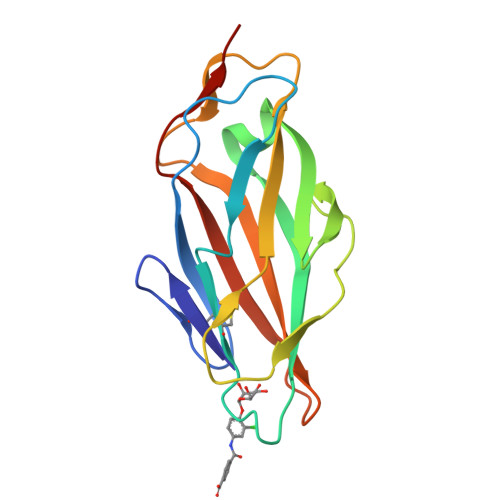
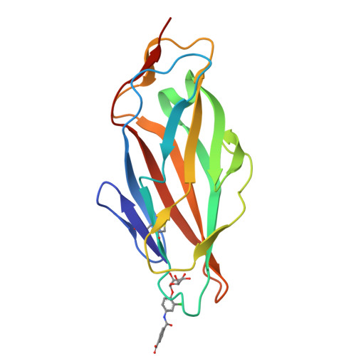
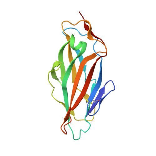
Crystal structure of FimH in complex with a benzoyl-amidophenyl alpha-D-mannopyranoside


Experimental Data Snapshot

  • Method: X-RAY DIFFRACTION
  • Resolution: 1.00 Å
  • R-Value Free: 0.139 
  • R-Value Work: 0.123 
  • R-Value Observed: 0.124 

Starting Model: experimental
View more details

wwPDB Validation   3D Report Full Report


Ligand Structure Quality Assessment 


This is version 1.3 of the entry. See complete history


Literature

The Tyrosine Gate of the Bacterial Lectin FimH: A Conformational Analysis by NMR Spectroscopy and X-ray Crystallography.

Fiege, B.Rabbani, S.Preston, R.C.Jakob, R.P.Zihlmann, P.Schwardt, O.Jiang, X.Maier, T.Ernst, B.

(2015) Chembiochem 16: 1235-1246

  • DOI: https://doi.org/10.1002/cbic.201402714
  • Primary Citation of Related Structures:  
    4X50, 4X5P, 4X5Q, 4X5R

  • PubMed Abstract: 

    Urinary tract infections caused by uropathogenic E. coli are among the most prevalent infectious diseases. The mannose-specific lectin FimH mediates the adhesion of the bacteria to the urothelium, thus enabling host cell invasion and recurrent infections. An attractive alternative to antibiotic treatment is the development of FimH antagonists that mimic the physiological ligand. A large variety of candidate drugs have been developed and characterized by means of in vitro studies and animal models. Here we present the X-ray co-crystal structures of FimH with members of four antagonist classes. In three of these cases no structural data had previously been available. We used NMR spectroscopy to characterize FimH-antagonist interactions further by chemical shift perturbation. The analysis allowed a clear determination of the conformation of the tyrosine gate motif that is crucial for the interaction with aglycone moieties and was not obvious from X-ray structural data alone. Finally, ITC experiments provided insight into the thermodynamics of antagonist binding. In conjunction with the structural information from X-ray and NMR experiments the results provide a mechanism for the often-observed enthalpy-entropy compensation of FimH antagonists that plays a role in fine-tuning of the interaction.


  • Organizational Affiliation

    Institute of Molecular Pharmacy, University of Basel, Klingelbergstrasse 50, 4056 Basel (Switzerland).


Macromolecules
Find similar proteins by:  (by identity cutoff)  |  3D Structure
Entity ID: 1
MoleculeChains Sequence LengthOrganismDetailsImage
Protein FimH160Escherichia coli K-12Mutation(s): 0 
Gene Names: fimHb4320JW4283
UniProt
Find proteins for P08191 (Escherichia coli (strain K12))
Explore P08191 
Go to UniProtKB:  P08191
Entity Groups  
Sequence Clusters30% Identity50% Identity70% Identity90% Identity95% Identity100% Identity
UniProt GroupP08191
Sequence Annotations
Expand
  • Reference Sequence
Small Molecules
Ligands 1 Unique
IDChains Name / Formula / InChI Key2D Diagram3D Interactions
3XJ
Query on 3XJ

Download Ideal Coordinates CCD File 
B [auth A]4-{[3-chloro-4-(alpha-D-mannopyranosyloxy)phenyl]carbamoyl}benzoic acid
C20 H20 Cl N O9
HIKPKPMJGGGESU-BAPGRXHISA-N
Experimental Data & Validation

Experimental Data

  • Method: X-RAY DIFFRACTION
  • Resolution: 1.00 Å
  • R-Value Free: 0.139 
  • R-Value Work: 0.123 
  • R-Value Observed: 0.124 
  • Space Group: P 21 21 21
Unit Cell:
Length ( Å )Angle ( ˚ )
a = 48.84α = 90
b = 55.89β = 90
c = 61γ = 90
Software Package:
Software NamePurpose
PHENIXrefinement
XDSdata reduction
XSCALEdata scaling
PHASERphasing

Structure Validation

View Full Validation Report


Ligand Structure Quality Assessment 


Entry History & Funding Information

Deposition Data


Funding OrganizationLocationGrant Number
Swiss National Science FoundationSwitzerland31003A_144183/1

Revision History  (Full details and data files)

  • Version 1.0: 2015-05-20
    Type: Initial release
  • Version 1.1: 2015-05-27
    Changes: Database references
  • Version 1.2: 2024-01-10
    Changes: Data collection, Database references, Refinement description
  • Version 1.3: 2024-11-13
    Changes: Structure summary