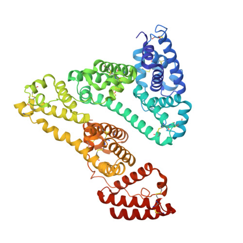
4Z69

Human serum albumin complexed with palmitic acid and diclofenac


Experimental Data Snapshot

  • Method: X-RAY DIFFRACTION
  • Resolution: 2.19 Å
  • R-Value Free: 0.293 
  • R-Value Work: 0.225 
  • R-Value Observed: 0.229 

wwPDB Validation   3D Report Full Report


Ligand Structure Quality Assessment 


This is version 1.1 of the entry. See complete history


Literature

Structural basis of non-steroidal anti-inflammatory drug diclofenac binding to human serum albumin.

Zhang, Y.Lee, P.Liang, S.Zhou, Z.Wu, X.Yang, F.Liang, H.

(2015) Chem Biol Drug Des 86: 1178-1184

  • DOI: https://doi.org/10.1111/cbdd.12583
  • Primary Citation of Related Structures:  
    4Z69

  • PubMed Abstract: 

    Human serum albumin (HSA) is the most abundant protein in plasma, which plays a central role in drug pharmacokinetics because most compounds bound to HSA in blood circulation. To understand binding characterization of non-steroidal anti-inflammatory drugs to HSA, we resolved the structure of diclofenac and HSA complex by X-ray crystallography. HSA-palmitic acid-diclofenac structure reveals two distinct binding sites for three diclofenac in HSA. One diclofenac is located at the IB subdomain, and its carboxylate group projects toward polar environment, forming hydrogen bond with one water molecule. The other two diclofenac molecules cobind in big hydrophobic cavity of the IIA subdomain without interactive association. Among them, one binds in main chamber of big hydrophobic cavity, and its carboxylate group forms hydrogen bonds with Lys199 and Arg218, as well as one water molecule, whereas another diclofenac binds in side chamber, its carboxylate group projects out cavity, forming hydrogen bond with Ser480.


  • Organizational Affiliation

    Key Laboratory of Ecology of Rare an Endangered Species and Environmental Protection, Ministry of Education of the People's Republic of China, Guangxi Normal University, Guilin, Guangxi, China.


Macromolecules
Find similar proteins by:  (by identity cutoff)  |  3D Structure
Entity ID: 1
MoleculeChains Sequence LengthOrganismDetailsImage
Serum albuminA,
B [auth I]
585Homo sapiensMutation(s): 0 
Gene Names: ALBGIG20GIG42PRO0903PRO1708PRO2044PRO2619PRO2675UNQ696/PRO1341
UniProt & NIH Common Fund Data Resources
Find proteins for P02768 (Homo sapiens)
Explore P02768 
Go to UniProtKB:  P02768
PHAROS:  P02768
GTEx:  ENSG00000163631 
Entity Groups  
Sequence Clusters30% Identity50% Identity70% Identity90% Identity95% Identity100% Identity
UniProt GroupP02768
Sequence Annotations
Expand
  • Reference Sequence
Small Molecules
Ligands 3 Unique
IDChains Name / Formula / InChI Key2D Diagram3D Interactions
DIF
Query on DIF

Download Ideal Coordinates CCD File 
H [auth A],
I [auth A],
J [auth A],
P [auth I]
2-[2,6-DICHLOROPHENYL)AMINO]BENZENEACETIC ACID
C14 H11 Cl2 N O2
DCOPUUMXTXDBNB-UHFFFAOYSA-N
PLM
Query on PLM

Download Ideal Coordinates CCD File 
D [auth A],
F [auth A],
L [auth I],
N [auth I]
PALMITIC ACID
C16 H32 O2
IPCSVZSSVZVIGE-UHFFFAOYSA-N
F15
Query on F15

Download Ideal Coordinates CCD File 
C [auth A]
E [auth A]
G [auth A]
K [auth I]
M [auth I]
C [auth A],
E [auth A],
G [auth A],
K [auth I],
M [auth I],
O [auth I]
PENTADECANOIC ACID
C15 H30 O2
WQEPLUUGTLDZJY-UHFFFAOYSA-N
Experimental Data & Validation

Experimental Data

  • Method: X-RAY DIFFRACTION
  • Resolution: 2.19 Å
  • R-Value Free: 0.293 
  • R-Value Work: 0.225 
  • R-Value Observed: 0.229 
  • Space Group: P 1
Unit Cell:
Length ( Å )Angle ( ˚ )
a = 38.396α = 105
b = 95.003β = 101.45
c = 96.339γ = 89.97
Software Package:
Software NamePurpose
DENZOdata collection
HKL-2000data scaling
PHENIXphasing
PHENIXrefinement

Structure Validation

View Full Validation Report


Ligand Structure Quality Assessment 


Entry History 

Deposition Data

Revision History  (Full details and data files)

  • Version 1.0: 2016-01-27
    Type: Initial release
  • Version 1.1: 2024-10-23
    Changes: Data collection, Database references, Derived calculations, Structure summary