4Z7V

L3-12 complex


Experimental Data Snapshot

  • Method: X-RAY DIFFRACTION
  • Resolution: 2.65 Å
  • R-Value Free: 
    0.226 (Depositor), 0.240 (DCC) 
  • R-Value Work: 
    0.187 (Depositor), 0.200 (DCC) 
  • R-Value Observed: 
    0.189 (Depositor) 

Starting Model: experimental
View more details

wwPDB Validation   3D Report Full Report


Ligand Structure Quality Assessment 

Created with Raphaël 2.3.0Worse 01 BetterLigand structure goodness of fit to experimental dataBest fitted NAGClick on this verticalbar to view details

This is version 2.2 of the entry. See complete history


Literature

Determinants of Gliadin-Specific T Cell Selection in Celiac Disease.

Petersen, J.van Bergen, J.Loh, K.L.Kooy-Winkelaar, Y.Beringer, D.X.Thompson, A.Bakker, S.F.Mulder, C.J.Ladell, K.McLaren, J.E.Price, D.A.Rossjohn, J.Reid, H.H.Koning, F.

(2015) J Immunol 194: 6112-6122

  • DOI: https://doi.org/10.4049/jimmunol.1500161
  • Primary Citation of Related Structures:  
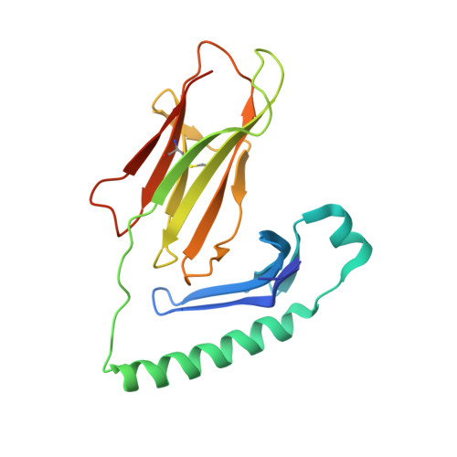
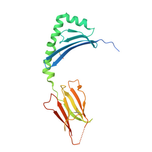
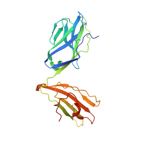
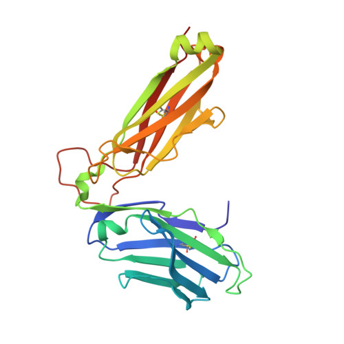
    4Z7U, 4Z7V, 4Z7W

  • PubMed Abstract: 

    In HLA-DQ8-associated celiac disease (CD), the pathogenic T cell response is directed toward an immunodominant α-gliadin-derived peptide (DQ8-glia-α1). However, our knowledge of TCR gene usage within the primary intestinal tissue of HLA-DQ8 (+) CD patients is limited. We identified two populations of HLA-DQ8-glia-α1 tetramer(+) CD4(+) T cells that were essentially undetectable in biopsy samples from patients on a gluten-free diet but expanded rapidly and specifically after antigenic stimulation. Distinguished by expression of TRBV9, both T cell populations displayed biased clonotypic repertoires and reacted similarly against HLA-DQ8-glia-α1. In particular, TRBV9 paired most often with TRAV26-2, whereas the majority of TRBV9(-) TCRs used TRBV6-1 with no clear TRAV gene preference. Strikingly, both tetramer(+)/TRBV9(+) and tetramer(+)/TRBV9(-) T cells possessed a non-germline-encoded arginine residue in their CDR3α and CDR3β loops, respectively. Comparison of the crystal structures of three TRBV9(+) TCRs and a TRBV9(-) TCR revealed that, as a result of distinct TCR docking modes, the HLA-DQ8-glia-α1 contacts mediated by the CDR3-encoded arginine were almost identical between TRBV9(+) and TRBV9(-) TCRs. In all cases, this interaction centered on two hydrogen bonds with a specific serine residue in the bound peptide. Replacement of serine with alanine at this position abrogated TRBV9(+) and TRBV9(-) clonal T cell proliferation in response to HLA-DQ8-glia-α1. Gluten-specific memory CD4(+) T cells with structurally and functionally conserved TCRs therefore predominate in the disease-affected tissue of patients with HLA-DQ8-mediated CD.


  • Organizational Affiliation

    Department of Biochemistry and Molecular Biology, School of Biomedical Sciences, Monash University, Clayton, Victoria 3800, Australia; Australian Research Council Centre of Excellence in Advanced Molecular Imaging, Monash University, Clayton, Victoria 3800, Australia;


Macromolecules
Find similar proteins by:  (by identity cutoff)  |  3D Structure
Entity ID: 1
MoleculeChains Sequence LengthOrganismDetailsImage
MHC class II HLA-DQ-alpha chain
A, C
192Homo sapiensMutation(s): 0 
Gene Names: HLA-DQA1
UniProt
Find proteins for Q30069 (Homo sapiens)
Explore Q30069 
Go to UniProtKB:  Q30069
Entity Groups  
Sequence Clusters30% Identity50% Identity70% Identity90% Identity95% Identity100% Identity
UniProt GroupQ30069
Glycosylation
Glycosylation Sites: 1
Sequence Annotations
Expand
  • Reference Sequence
Find similar proteins by:  (by identity cutoff)  |  3D Structure
Entity ID: 2
MoleculeChains Sequence LengthOrganismDetailsImage
MHC class II HLA-DQ-beta-1
B, D
213Homo sapiensMutation(s): 0 
Gene Names: HLA-DQB1
UniProt
Find proteins for O19707 (Homo sapiens)
Explore O19707 
Go to UniProtKB:  O19707
Entity Groups  
Sequence Clusters30% Identity50% Identity70% Identity90% Identity95% Identity100% Identity
UniProt GroupO19707
Glycosylation
Glycosylation Sites: 1
Sequence Annotations
Expand
  • Reference Sequence
Find similar proteins by:  (by identity cutoff)  |  3D Structure
Entity ID: 3
MoleculeChains Sequence LengthOrganismDetailsImage
T-CELL RECEPTOR, L3-12 ALPHA CHAIN
E, G
204Homo sapiensMutation(s): 0 
Entity Groups  
Sequence Clusters30% Identity50% Identity70% Identity90% Identity95% Identity100% Identity
Sequence Annotations
Expand
  • Reference Sequence
Find similar proteins by:  (by identity cutoff)  |  3D Structure
Entity ID: 4
MoleculeChains Sequence LengthOrganismDetailsImage
T-CELL RECEPTOR, L3-12 BETA CHAIN
F, H
244Homo sapiensMutation(s): 0 
Entity Groups  
Sequence Clusters30% Identity50% Identity70% Identity90% Identity95% Identity100% Identity
Sequence Annotations
Expand
  • Reference Sequence

Find similar proteins by:  Sequence   |   3D Structure  

Entity ID: 5
MoleculeChains Sequence LengthOrganismDetailsImage
deamidated DQ8-glia-alpha1 peptide
I, J
18Triticum aestivumMutation(s): 0 
UniProt
Find proteins for P18573 (Triticum aestivum)
Explore P18573 
Go to UniProtKB:  P18573
Entity Groups  
Sequence Clusters30% Identity50% Identity70% Identity90% Identity95% Identity100% Identity
UniProt GroupP18573
Sequence Annotations
Expand
  • Reference Sequence
Oligosaccharides

Help

Entity ID: 6
MoleculeChains Length2D Diagram Glycosylation3D Interactions
alpha-D-mannopyranose-(1-4)-alpha-D-mannopyranose-(1-4)-2-acetamido-2-deoxy-beta-D-glucopyranose-(1-4)-[alpha-L-fucopyranose-(1-3)][alpha-L-fucopyranose-(1-6)]2-acetamido-2-deoxy-beta-D-glucopyranose
K
6N-Glycosylation
Glycosylation Resources
GlyTouCan:  G73409CU
GlyCosmos:  G73409CU
GlyGen:  G73409CU
Entity ID: 7
MoleculeChains Length2D Diagram Glycosylation3D Interactions
alpha-L-fucopyranose-(1-3)-[2-acetamido-2-deoxy-beta-D-glucopyranose-(1-4)][alpha-L-fucopyranose-(1-6)]2-acetamido-2-deoxy-beta-D-glucopyranose
L
4N-Glycosylation
Glycosylation Resources
GlyTouCan:  G63564LA
GlyCosmos:  G63564LA
Experimental Data & Validation

Experimental Data

  • Method: X-RAY DIFFRACTION
  • Resolution: 2.65 Å
  • R-Value Free:  0.226 (Depositor), 0.240 (DCC) 
  • R-Value Work:  0.187 (Depositor), 0.200 (DCC) 
  • R-Value Observed: 0.189 (Depositor) 
Space Group: P 1
Unit Cell:
Length ( Å )Angle ( ˚ )
a = 56.51α = 93.87
b = 76.961β = 89.47
c = 132.279γ = 105.09
Software Package:
Software NamePurpose
BUSTERrefinement
XDSdata reduction
SCALAdata scaling
PHASERphasing

Structure Validation

View Full Validation Report


Ligand Structure Quality Assessment 

Created with Raphaël 2.3.0Worse 01 BetterLigand structure goodness of fit to experimental dataBest fitted NAGClick on this verticalbar to view details

Entry History 

Deposition Data

Revision History  (Full details and data files)

  • Version 1.0: 2015-06-03
    Type: Initial release
  • Version 1.1: 2015-06-17
    Changes: Database references
  • Version 1.2: 2017-08-23
    Changes: Data collection, Derived calculations, Source and taxonomy
  • Version 2.0: 2020-07-29
    Type: Remediation
    Reason: Carbohydrate remediation
    Changes: Advisory, Atomic model, Data collection, Derived calculations, Structure summary
  • Version 2.1: 2023-09-27
    Changes: Data collection, Database references, Derived calculations, Refinement description, Structure summary
  • Version 2.2: 2024-11-20
    Changes: Structure summary