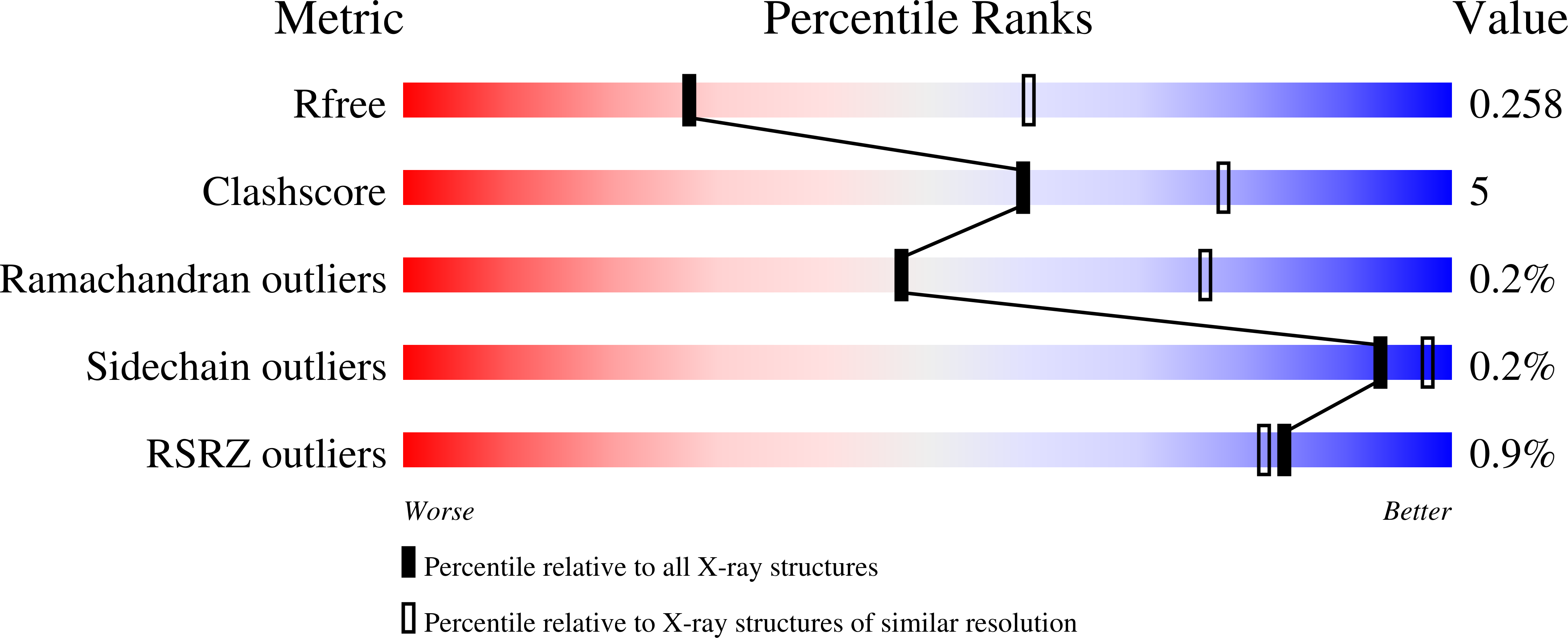
4ZF6

Cytochrome P450 pentamutant from BM3 with bound PEG


Experimental Data Snapshot

  • Method: X-RAY DIFFRACTION
  • Resolution: 2.77 Å
  • R-Value Free: 0.257 
  • R-Value Work: 0.201 
  • R-Value Observed: 0.204 

Starting Model: experimental
View more details

wwPDB Validation   3D Report Full Report


Ligand Structure Quality Assessment 


This is version 1.1 of the entry. See complete history


Literature

Effect of Mutation and Substrate Binding on the Stability of Cytochrome P450BM3 Variants.

Geronimo, I.Denning, C.A.Rogers, W.E.Othman, T.Huxford, T.Heidary, D.K.Glazer, E.C.Payne, C.M.

(2016) Biochemistry 55: 3594-3606

  • DOI: https://doi.org/10.1021/acs.biochem.6b00183
  • Primary Citation of Related Structures:  
    4ZF6, 4ZF8, 4ZFA, 4ZFB

  • PubMed Abstract: 

    Cytochrome P450BM3 is a heme-containing enzyme from Bacillus megaterium that exhibits high monooxygenase activity and has a self-sufficient electron transfer system in the full-length enzyme. Its potential synthetic applications drive protein engineering efforts to produce variants capable of oxidizing nonnative substrates such as pharmaceuticals and aromatic pollutants. However, promiscuous P450BM3 mutants often exhibit lower stability, thereby hindering their industrial application. This study demonstrated that the heme domain R47L/F87V/L188Q/E267V/F81I pentuple mutant (PM) is destabilized because of the disruption of hydrophobic contacts and salt bridge interactions. This was directly observed from crystal structures of PM in the presence and absence of ligands (palmitic acid and metyrapone). The instability of the tertiary structure and heme environment of substrate-free PM was confirmed by pulse proteolysis and circular dichroism, respectively. Binding of the inhibitor, metyrapone, significantly stabilized PM, but the presence of the native substrate, palmitic acid, had no effect. On the basis of high-temperature molecular dynamics simulations, the lid domain, β-sheet 1, and Cys ligand loop (a β-bulge segment connected to the heme) are the most labile regions and, thus, potential sites for stabilizing mutations. Possible approaches to stabilization include improvement of hydrophobic packing interactions in the lid domain and introduction of new salt bridges into β-sheet 1 and the heme region. An understanding of the molecular factors behind the loss of stability of P450BM3 variants therefore expedites site-directed mutagenesis studies aimed at developing thermostability.


  • Organizational Affiliation

    Department of Chemical and Materials Engineering, University of Kentucky , Lexington, Kentucky 40506-0046, United States.


Macromolecules
Find similar proteins by:  (by identity cutoff)  |  3D Structure
Entity ID: 1
MoleculeChains Sequence LengthOrganismDetailsImage
Bifunctional P-450/NADPH-P450 reductase461Priestia megateriumMutation(s): 5 
Gene Names: cyp102A1cyp102
EC: 1.14.14.1 (PDB Primary Data), 1.6.2.4 (PDB Primary Data)
UniProt
Find proteins for P14779 (Priestia megaterium (strain ATCC 14581 / DSM 32 / CCUG 1817 / JCM 2506 / NBRC 15308 / NCIMB 9376 / NCTC 10342 / NRRL B-14308 / VKM B-512 / Ford 19))
Explore P14779 
Go to UniProtKB:  P14779
Entity Groups  
Sequence Clusters30% Identity50% Identity70% Identity90% Identity95% Identity100% Identity
UniProt GroupP14779
Sequence Annotations
Expand
  • Reference Sequence
Small Molecules
Ligands 4 Unique
IDChains Name / Formula / InChI Key2D Diagram3D Interactions
HEM
Query on HEM

Download Ideal Coordinates CCD File 
B [auth A]PROTOPORPHYRIN IX CONTAINING FE
C34 H32 Fe N4 O4
KABFMIBPWCXCRK-RGGAHWMASA-L
1PE
Query on 1PE

Download Ideal Coordinates CCD File 
C [auth A]PENTAETHYLENE GLYCOL
C10 H22 O6
JLFNLZLINWHATN-UHFFFAOYSA-N
EDO
Query on EDO

Download Ideal Coordinates CCD File 
D [auth A]1,2-ETHANEDIOL
C2 H6 O2
LYCAIKOWRPUZTN-UHFFFAOYSA-N
NI
Query on NI

Download Ideal Coordinates CCD File 
E [auth A],
F [auth A],
G [auth A],
H [auth A]
NICKEL (II) ION
Ni
VEQPNABPJHWNSG-UHFFFAOYSA-N
Experimental Data & Validation

Experimental Data

  • Method: X-RAY DIFFRACTION
  • Resolution: 2.77 Å
  • R-Value Free: 0.257 
  • R-Value Work: 0.201 
  • R-Value Observed: 0.204 
  • Space Group: P 65 2 2
Unit Cell:
Length ( Å )Angle ( ˚ )
a = 56.047α = 90
b = 56.047β = 90
c = 711.229γ = 120
Software Package:
Software NamePurpose
PHENIXrefinement
Cootmodel building
PHENIXphasing
XDSdata reduction
XDSdata scaling

Structure Validation

View Full Validation Report


Ligand Structure Quality Assessment 


Entry History 

Deposition Data

Revision History  (Full details and data files)

  • Version 1.0: 2016-07-13
    Type: Initial release
  • Version 1.1: 2023-09-27
    Changes: Data collection, Database references, Derived calculations, Refinement description