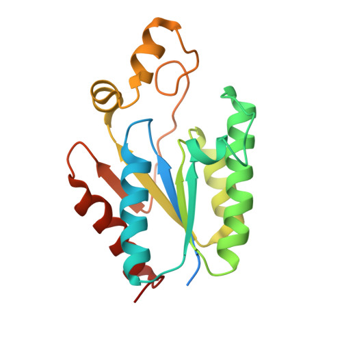
5CB6

Structure of adenosine-5'-phosphosulfate kinase


Experimental Data Snapshot

  • Method: X-RAY DIFFRACTION
  • Resolution: 2.79 Å
  • R-Value Free: 0.218 
  • R-Value Work: 0.164 
  • R-Value Observed: 0.167 

Starting Model: experimental
View more details

wwPDB Validation   3D Report Full Report


Ligand Structure Quality Assessment 


This is version 1.5 of the entry. See complete history


Literature

Recapitulating the Structural Evolution of Redox Regulation in Adenosine 5'-Phosphosulfate Kinase from Cyanobacteria to Plants.

Herrmann, J.Nathin, D.Lee, S.G.Sun, T.Jez, J.M.

(2015) J Biol Chem 290: 24705-24714

  • DOI: https://doi.org/10.1074/jbc.M115.679514
  • Primary Citation of Related Structures:  
    5CB6, 5CB8

  • PubMed Abstract: 

    In plants, adenosine 5'-phosphosulfate (APS) kinase (APSK) is required for reproductive viability and the production of 3'-phosphoadenosine 5'-phosphosulfate (PAPS) as a sulfur donor in specialized metabolism. Previous studies of the APSK from Arabidopsis thaliana (AtAPSK) identified a regulatory disulfide bond formed between the N-terminal domain (NTD) and a cysteine on the core scaffold. This thiol switch is unique to mosses, gymnosperms, and angiosperms. To understand the structural evolution of redox control of APSK, we investigated the redox-insensitive APSK from the cyanobacterium Synechocystis sp. PCC 6803 (SynAPSK). Crystallographic analysis of SynAPSK in complex with either APS and a non-hydrolyzable ATP analog or APS and sulfate revealed the overall structure of the enzyme, which lacks the NTD found in homologs from mosses and plants. A series of engineered SynAPSK variants reconstructed the structural evolution of the plant APSK. Biochemical analyses of SynAPSK, SynAPSK H23C mutant, SynAPSK fused to the AtAPSK NTD, and the fusion protein with the H23C mutation showed that the addition of the NTD and cysteines recapitulated thiol-based regulation. These results reveal the molecular basis for structural changes leading to the evolution of redox control of APSK in the green lineage from cyanobacteria to plants.


  • Organizational Affiliation

    From the Department of Biology, Washington University, St. Louis, Missouri 63130.


Macromolecules
Find similar proteins by:  (by identity cutoff)  |  3D Structure
Entity ID: 1
MoleculeChains Sequence LengthOrganismDetailsImage
Probable adenylyl-sulfate kinase
A, B, C, D, E
A, B, C, D, E, F
197Synechocystis sp. PCC 6803 substr. KazusaMutation(s): 0 
Gene Names: cysCslr0676
EC: 2.7.1.25
UniProt
Find proteins for P72940 (Synechocystis sp. (strain ATCC 27184 / PCC 6803 / Kazusa))
Explore P72940 
Go to UniProtKB:  P72940
Entity Groups  
Sequence Clusters30% Identity50% Identity70% Identity90% Identity95% Identity100% Identity
UniProt GroupP72940
Sequence Annotations
Expand
  • Reference Sequence
Small Molecules
Ligands 5 Unique
IDChains Name / Formula / InChI Key2D Diagram3D Interactions
ANP
Query on ANP

Download Ideal Coordinates CCD File 
H [auth A]
L [auth B]
O [auth C]
S [auth D]
W [auth E]
H [auth A],
L [auth B],
O [auth C],
S [auth D],
W [auth E],
Z [auth F]
PHOSPHOAMINOPHOSPHONIC ACID-ADENYLATE ESTER
C10 H17 N6 O12 P3
PVKSNHVPLWYQGJ-KQYNXXCUSA-N
ADX
Query on ADX

Download Ideal Coordinates CCD File 
G [auth A]
K [auth B]
N [auth C]
R [auth D]
V [auth E]
G [auth A],
K [auth B],
N [auth C],
R [auth D],
V [auth E],
Y [auth F]
ADENOSINE-5'-PHOSPHOSULFATE
C10 H14 N5 O10 P S
IRLPACMLTUPBCL-KQYNXXCUSA-N
CAC
Query on CAC

Download Ideal Coordinates CCD File 
Q [auth C],
U [auth D]
CACODYLATE ION
C2 H6 As O2
OGGXGZAMXPVRFZ-UHFFFAOYSA-M
MG
Query on MG

Download Ideal Coordinates CCD File 
AA [auth F]
I [auth A]
M [auth B]
P [auth C]
T [auth D]
AA [auth F],
I [auth A],
M [auth B],
P [auth C],
T [auth D],
X [auth E]
MAGNESIUM ION
Mg
JLVVSXFLKOJNIY-UHFFFAOYSA-N
NA
Query on NA

Download Ideal Coordinates CCD File 
J [auth A]SODIUM ION
Na
FKNQFGJONOIPTF-UHFFFAOYSA-N
Experimental Data & Validation

Experimental Data

  • Method: X-RAY DIFFRACTION
  • Resolution: 2.79 Å
  • R-Value Free: 0.218 
  • R-Value Work: 0.164 
  • R-Value Observed: 0.167 
  • Space Group: C 1 2 1
Unit Cell:
Length ( Å )Angle ( ˚ )
a = 136.904α = 90
b = 109.681β = 90.72
c = 100.807γ = 90
Software Package:
Software NamePurpose
PHENIXrefinement
HKL-3000data reduction
HKL-3000data scaling
PHENIXphasing

Structure Validation

View Full Validation Report


Ligand Structure Quality Assessment 


Entry History & Funding Information

Deposition Data


Funding OrganizationLocationGrant Number
National Science Foundation (NSF, United States)United StatesMCB-0904215

Revision History  (Full details and data files)

  • Version 1.0: 2015-08-26
    Type: Initial release
  • Version 1.1: 2015-09-02
    Changes: Database references
  • Version 1.2: 2015-10-21
    Changes: Database references
  • Version 1.3: 2017-09-27
    Changes: Author supporting evidence, Database references, Derived calculations
  • Version 1.4: 2019-11-27
    Changes: Author supporting evidence
  • Version 1.5: 2023-09-27
    Changes: Data collection, Database references, Derived calculations, Refinement description