5EBV

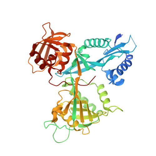
Crystal structure of acetyltransferase Eis from Mycobacterium tuberculosis in complex with inhibitor 11c and CoA


Experimental Data Snapshot

  • Method: X-RAY DIFFRACTION
  • Resolution: 2.20 Å
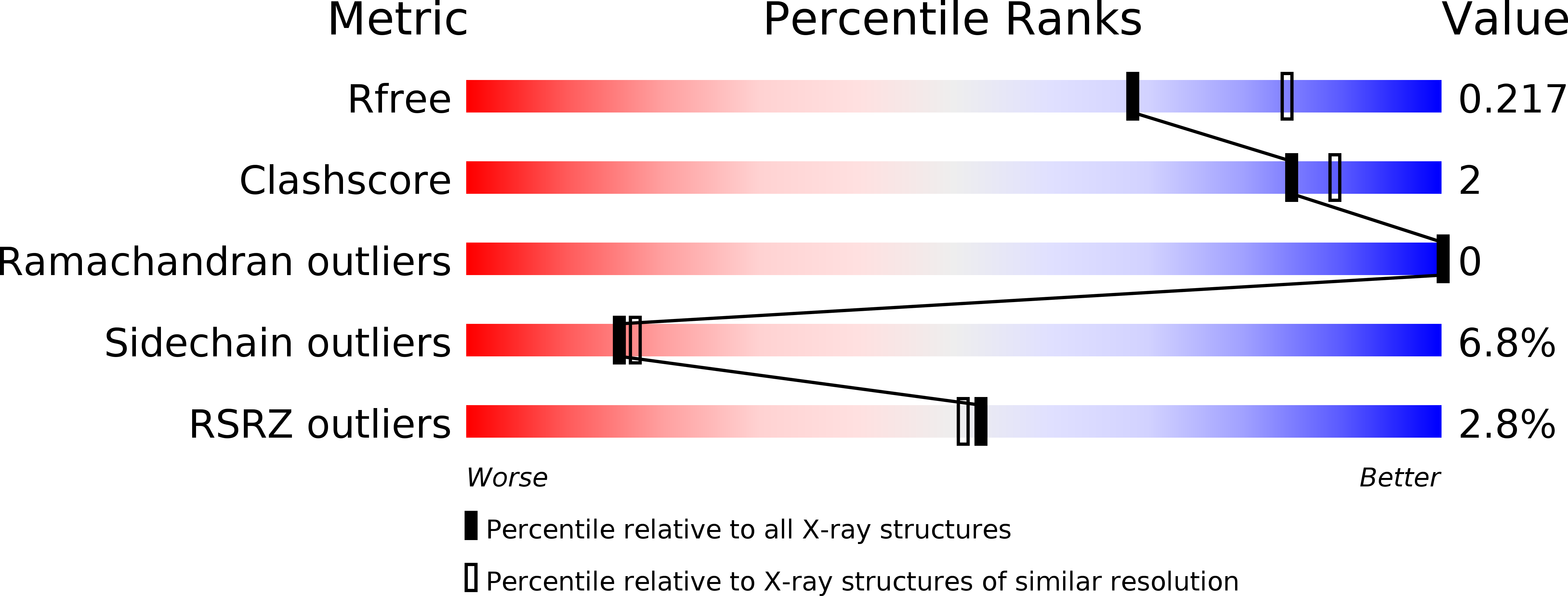
  • R-Value Free: 0.215 
  • R-Value Work: 0.193 
  • R-Value Observed: 0.194 

wwPDB Validation   3D Report Full Report


Ligand Structure Quality Assessment 


This is version 1.5 of the entry. See complete history


Literature

Potent Inhibitors of Acetyltransferase Eis Overcome Kanamycin Resistance in Mycobacterium tuberculosis.

Willby, M.J.Green, K.D.Gajadeera, C.S.Hou, C.Tsodikov, O.V.Posey, J.E.Garneau-Tsodikova, S.

(2016) ACS Chem Biol 11: 1639-1646

  • DOI: https://doi.org/10.1021/acschembio.6b00110
  • Primary Citation of Related Structures:  
    5EBV, 5EC4

  • PubMed Abstract: 

    A major cause of tuberculosis (TB) resistance to the aminoglycoside kanamycin (KAN) is the Mycobacterium tuberculosis (Mtb) acetyltransferase Eis. Upregulation of this enzyme is responsible for inactivation of KAN through acetylation of its amino groups. A 123 000-compound high-throughput screen (HTS) yielded several small-molecule Eis inhibitors that share an isothiazole S,S-dioxide heterocyclic core. These were investigated for their structure-activity relationships. Crystal structures of Eis in complex with two potent inhibitors show that these molecules are bound in the conformationally adaptable aminoglycoside binding site of the enzyme, thereby obstructing binding of KAN for acetylation. Importantly, we demonstrate that several Eis inhibitors, when used in combination with KAN against resistant Mtb, efficiently overcome KAN resistance. This approach paves the way toward development of novel combination therapies against aminoglycoside-resistant TB.


  • Organizational Affiliation

    Division of Tuberculosis Elimination, National Center for HIV/AIDS, Viral Hepatitis, STD, and TB Prevention, Centers for Disease Control and Prevention , Atlanta, Georgia 30329, United States.


Macromolecules
Find similar proteins by:  (by identity cutoff)  |  3D Structure
Entity ID: 1
MoleculeChains Sequence LengthOrganismDetailsImage
Enhanced intracellular survival protein422Mycobacterium tuberculosis H37RvMutation(s): 1 
Gene Names: eisRv2416cMTCY253.04
EC: 2.3.1
UniProt
Find proteins for P9WFK7 (Mycobacterium tuberculosis (strain ATCC 25618 / H37Rv))
Explore P9WFK7 
Go to UniProtKB:  P9WFK7
Entity Groups  
Sequence Clusters30% Identity50% Identity70% Identity90% Identity95% Identity100% Identity
UniProt GroupP9WFK7
Sequence Annotations
Expand
  • Reference Sequence
Small Molecules
Ligands 3 Unique
IDChains Name / Formula / InChI Key2D Diagram3D Interactions
COA
Query on COA

Download Ideal Coordinates CCD File 
B [auth A]COENZYME A
C21 H36 N7 O16 P3 S
RGJOEKWQDUBAIZ-IBOSZNHHSA-N
5LR
Query on 5LR

Download Ideal Coordinates CCD File 
C [auth A]5-(4-chlorophenyl)-~{N}-[3-(3,4-dihydro-1~{H}-isoquinolin-2-yl)propyl]-4-methyl-1,1-bis(oxidanylidene)-1,2-thiazol-3-amine
C22 H24 Cl N3 O2 S
MAOQHBNJMHYTOS-UHFFFAOYSA-N
CL
Query on CL

Download Ideal Coordinates CCD File 
D [auth A]CHLORIDE ION
Cl
VEXZGXHMUGYJMC-UHFFFAOYSA-M
Experimental Data & Validation

Experimental Data

  • Method: X-RAY DIFFRACTION
  • Resolution: 2.20 Å
  • R-Value Free: 0.215 
  • R-Value Work: 0.193 
  • R-Value Observed: 0.194 
  • Space Group: H 3 2
Unit Cell:
Length ( Å )Angle ( ˚ )
a = 175.168α = 90
b = 175.168β = 90
c = 122.32γ = 120
Software Package:
Software NamePurpose
REFMACrefinement
HKL-2000data reduction
HKL-2000data scaling
PHASERphasing

Structure Validation

View Full Validation Report


Ligand Structure Quality Assessment 


Entry History & Funding Information

Deposition Data


Funding OrganizationLocationGrant Number
National Institutes of Health/National Institute Of Allergy and Infectious Diseases (NIH/NIAID)United StatesAI090048

Revision History  (Full details and data files)

  • Version 1.0: 2016-03-30
    Type: Initial release
  • Version 1.1: 2016-04-06
    Changes: Database references
  • Version 1.2: 2016-06-29
    Changes: Database references
  • Version 1.3: 2017-09-20
    Changes: Author supporting evidence, Derived calculations
  • Version 1.4: 2019-12-11
    Changes: Author supporting evidence
  • Version 1.5: 2024-03-06
    Changes: Data collection, Database references