5EFW | pdb_00005efw

Crystal structure of LOV2-Zdk1 - the complex of oat LOV2 and the affibody protein Zdark1


Experimental Data Snapshot

  • Method: X-RAY DIFFRACTION
  • Resolution: 2.10 Å
  • R-Value Free: 
    0.257 (Depositor), 0.260 (DCC) 
  • R-Value Work: 
    0.202 (Depositor), 0.200 (DCC) 
  • R-Value Observed: 
    0.204 (Depositor) 

Starting Models: experimental
View more details

wwPDB Validation   3D Report Full Report


Ligand Structure Quality Assessment 

Created with Raphaël 2.3.0Worse 01 BetterLigand structure goodness of fit to experimental dataBest fitted FMNClick on this verticalbar to view details

This is version 1.4 of the entry. See complete history


Literature

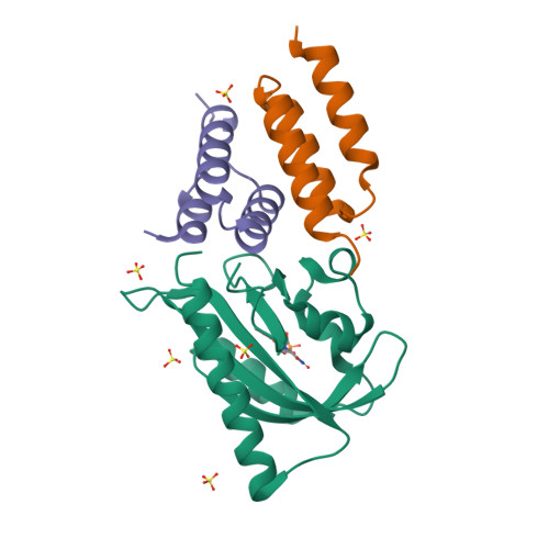
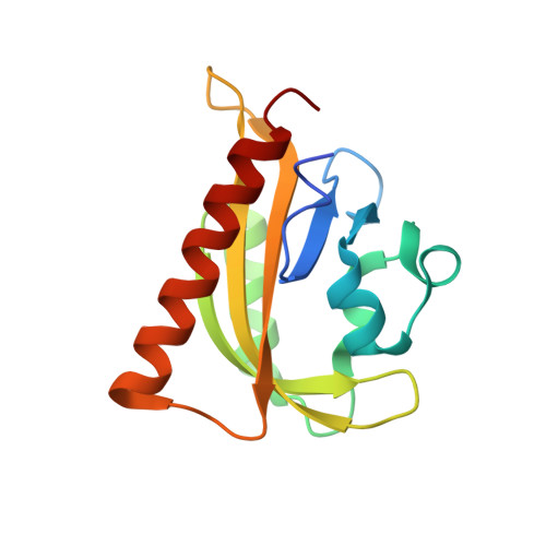
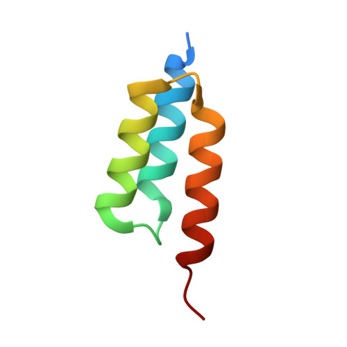
LOVTRAP: an optogenetic system for photoinduced protein dissociation.

Wang, H.Vilela, M.Winkler, A.Tarnawski, M.Schlichting, I.Yumerefendi, H.Kuhlman, B.Liu, R.Danuser, G.Hahn, K.M.

(2016) Nat Methods 13: 755-758

  • DOI: https://doi.org/10.1038/nmeth.3926
  • Primary Citation of Related Structures:  
    5DJT, 5DJU, 5EFW

  • PubMed Abstract: 

    LOVTRAP is an optogenetic approach for reversible light-induced protein dissociation using protein A fragments that bind to the LOV domain only in the dark, with tunable kinetics and a >150-fold change in the dissociation constant (Kd). By reversibly sequestering proteins at mitochondria, we precisely modulated the proteins' access to the cell edge, demonstrating a naturally occurring 3-mHz cell-edge oscillation driven by interactions of Vav2, Rac1, and PI3K proteins.


  • Organizational Affiliation

    Department of Pharmacology, University of North Carolina at Chapel Hill, Chapel Hill, North Carolina, USA.


Macromolecules
Find similar proteins by:  (by identity cutoff)  |  3D Structure
Entity ID: 1
MoleculeChains Sequence LengthOrganismDetailsImage
NPH1-1145Avena sativaMutation(s): 1 
Gene Names: NPH1-1
EC: 2.7.11.1
UniProt
Find proteins for O49003 (Avena sativa)
Explore O49003 
Go to UniProtKB:  O49003
Entity Groups  
Sequence Clusters30% Identity50% Identity70% Identity90% Identity95% Identity100% Identity
UniProt GroupO49003
Sequence Annotations
Expand
  • Reference Sequence
Find similar proteins by:  (by identity cutoff)  |  3D Structure
Entity ID: 2
MoleculeChains Sequence LengthOrganismDetailsImage
Z-dark, a small protein based on the Z domain affibody
B, C
60Staphylococcus aureusMutation(s): 0 
Entity Groups  
Sequence Clusters30% Identity50% Identity70% Identity90% Identity95% Identity100% Identity
Sequence Annotations
Expand
  • Reference Sequence
Experimental Data & Validation

Experimental Data

  • Method: X-RAY DIFFRACTION
  • Resolution: 2.10 Å
  • R-Value Free:  0.257 (Depositor), 0.260 (DCC) 
  • R-Value Work:  0.202 (Depositor), 0.200 (DCC) 
  • R-Value Observed: 0.204 (Depositor) 
Space Group: P 41 21 2
Unit Cell:
Length ( Å )Angle ( ˚ )
a = 54.7α = 90
b = 54.7β = 90
c = 188.03γ = 90
Software Package:
Software NamePurpose
XDSdata reduction
Cootmodel building
PHENIXrefinement
PDB_EXTRACTdata extraction
XSCALEdata scaling
PHASERphasing
XSCALEdata reduction

Structure Validation

View Full Validation Report


Ligand Structure Quality Assessment 

Created with Raphaël 2.3.0Worse 01 BetterLigand structure goodness of fit to experimental dataBest fitted FMNClick on this verticalbar to view details

Entry History & Funding Information

Deposition Data


Funding OrganizationLocationGrant Number
German Research FoundationGermanyFOR1279

Revision History  (Full details and data files)

  • Version 1.0: 2016-07-20
    Type: Initial release
  • Version 1.1: 2016-07-27
    Changes: Database references
  • Version 1.2: 2016-09-07
    Changes: Database references
  • Version 1.3: 2018-01-31
    Changes: Author supporting evidence, Database references
  • Version 1.4: 2024-01-10
    Changes: Data collection, Database references, Refinement description