5J5G

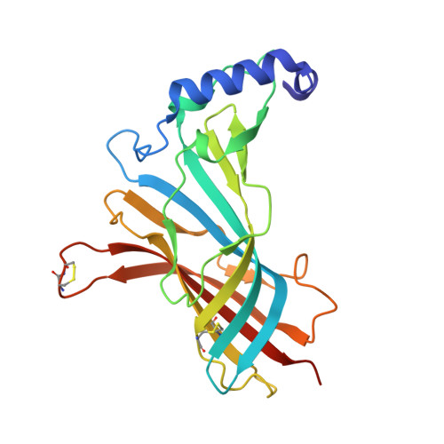
X-Ray Crystal Structure of Acetylcholine Binding Protein (AChBP) in Complex with 6-(4-methoxyphenyl)-N4,N4-bis[(pyridin-2-yl)methyl]pyrimidine-2,4-diamine


Experimental Data Snapshot

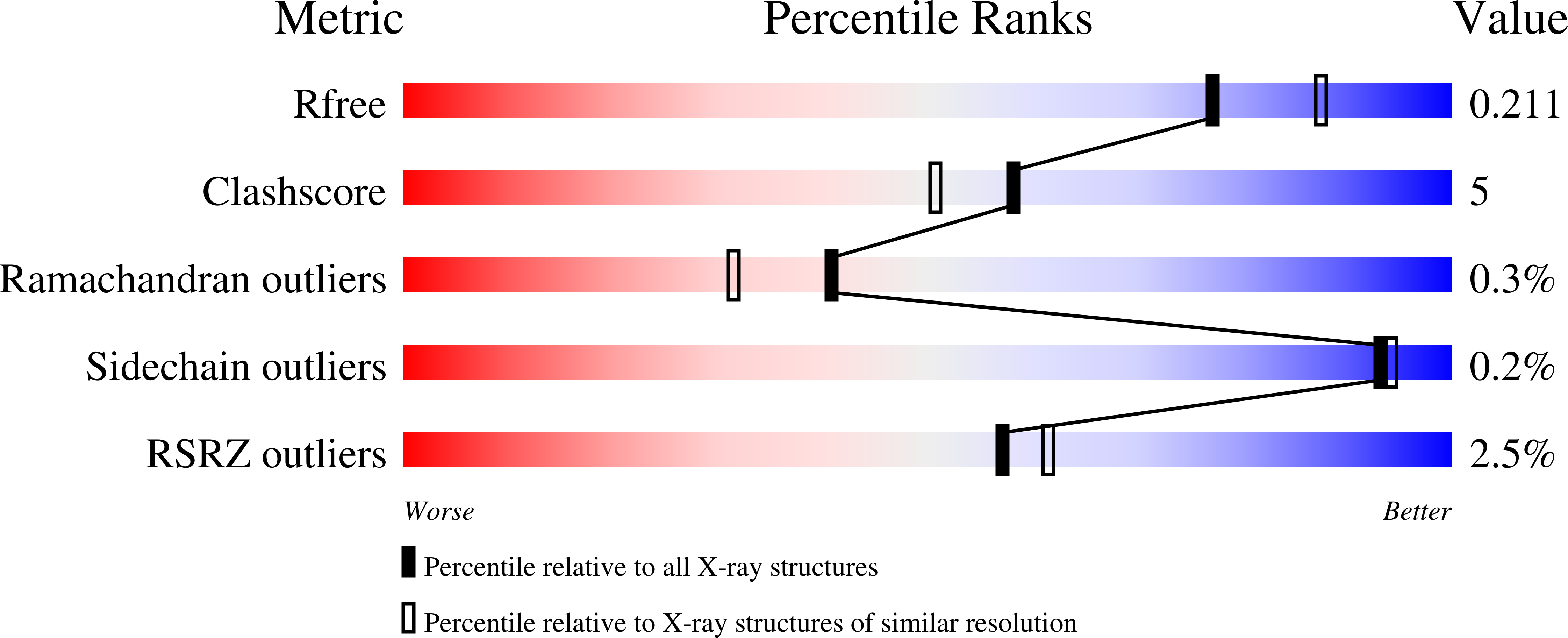
  • Method: X-RAY DIFFRACTION
  • Resolution: 2.04 Å
  • R-Value Free: 0.209 
  • R-Value Work: 0.166 
  • R-Value Observed: 0.167 

Starting Model: experimental
View more details

wwPDB Validation   3D Report Full Report


Ligand Structure Quality Assessment 


This is version 1.5 of the entry. See complete history


Literature

Substituted 2-Aminopyrimidines Selective for alpha 7-Nicotinic Acetylcholine Receptor Activation and Association with Acetylcholine Binding Proteins.

Kaczanowska, K.Camacho Hernandez, G.A.Bendiks, L.Kohs, L.Cornejo-Bravo, J.M.Harel, M.Finn, M.G.Taylor, P.

(2017) J Am Chem Soc 139: 3676-3684

  • DOI: https://doi.org/10.1021/jacs.6b10746
  • Primary Citation of Related Structures:  
    5J5F, 5J5G, 5J5H, 5J5I

  • PubMed Abstract: 

    Through studies with ligand binding to the acetylcholine binding protein (AChBP), we previously identified a series of 4,6-substituted 2-aminopyrimidines that associate with this soluble surrogate of the nicotinic acetylcholine receptor (nAChR) in a cooperative fashion, not seen for classical nicotinic agonists and antagonists. To examine receptor interactions of this structural family on ligand-gated ion channels, we employed HEK cells transfected with cDNAs encoding three requisite receptor subtypes: α7-nAChR, α4β2-nAChR, and a serotonin receptor (5-HT 3A R), along with a fluorescent reporter. Initial screening of a series of over 50 newly characterized 2-aminopyrimidines with affinity for AChBP showed only two to be agonists on the α7-nAChR below 10 μM concentration. Their unique structural features were incorporated into design of a second subset of 2-aminopyrimidines yielding several congeners that elicited α7 activation with EC 50 values of 70 nM and K d values for AChBP in a similar range. Several compounds within this series exhibit specificity for the α7-nAChR, showing no activation or antagonism of α4β2-nAChR or 5-HT3AR at concentrations up to 10 μM, while others were weaker antagonists (or partial agonists) on these receptors. Analysis following cocrystallization of four ligand complexes with AChBP show binding at the subunit interface, but with an orientation or binding pose that differs from classical nicotinic agonists and antagonists and from the previously analyzed set of 2-aminopyrimidines that displayed distinct cooperative interactions with AChBP. Orientations of aromatic side chains of these complexes are distinctive, suggesting new modes of binding at the agonist-antagonist site and perhaps an allosteric action for heteromeric nAChRs.


  • Organizational Affiliation

    Department of Pharmacology, Skaggs School of Pharmacy & Pharmaceutical Sciences, University of California, San Diego , La Jolla, California 92093-0650, United States.


Macromolecules
Find similar proteins by:  (by identity cutoff)  |  3D Structure
Entity ID: 1
MoleculeChains Sequence LengthOrganismDetailsImage
Acetylcholine-binding protein
A, B, C, D, E
A, B, C, D, E, F, G, H, I, J
218Lymnaea stagnalisMutation(s): 0 
UniProt
Find proteins for P58154 (Lymnaea stagnalis)
Explore P58154 
Go to UniProtKB:  P58154
Entity Groups  
Sequence Clusters30% Identity50% Identity70% Identity90% Identity95% Identity100% Identity
UniProt GroupP58154
Glycosylation
Glycosylation Sites: 1
Sequence Annotations
Expand
  • Reference Sequence
Small Molecules
Ligands 3 Unique
IDChains Name / Formula / InChI Key2D Diagram3D Interactions
6GF
Query on 6GF

Download Ideal Coordinates CCD File 
AA [auth F]
BA [auth F]
HA [auth H]
JA [auth I]
N [auth A]
AA [auth F],
BA [auth F],
HA [auth H],
JA [auth I],
N [auth A],
NA [auth J],
S [auth C],
T [auth C],
X [auth E],
Y [auth E]
6-(4-methoxyphenyl)-N~4~,N~4~-bis[(pyridin-2-yl)methyl]pyrimidine-2,4-diamine
C23 H22 N6 O
FIGQTBBMNODYCB-UHFFFAOYSA-N
NAG
Query on NAG

Download Ideal Coordinates CCD File 
DA [auth G]
GA [auth H]
IA [auth I]
M [auth A]
MA [auth J]
DA [auth G],
GA [auth H],
IA [auth I],
M [auth A],
MA [auth J],
O [auth B],
R [auth C],
U [auth D],
W [auth E],
Z [auth F]
2-acetamido-2-deoxy-beta-D-glucopyranose
C8 H15 N O6
OVRNDRQMDRJTHS-FMDGEEDCSA-N
PO4
Query on PO4

Download Ideal Coordinates CCD File 
CA [auth G]
EA [auth H]
FA [auth H]
K [auth A]
KA [auth J]
CA [auth G],
EA [auth H],
FA [auth H],
K [auth A],
KA [auth J],
L [auth A],
LA [auth J],
P [auth C],
Q [auth C],
V [auth E]
PHOSPHATE ION
O4 P
NBIIXXVUZAFLBC-UHFFFAOYSA-K
Experimental Data & Validation

Experimental Data

  • Method: X-RAY DIFFRACTION
  • Resolution: 2.04 Å
  • R-Value Free: 0.209 
  • R-Value Work: 0.166 
  • R-Value Observed: 0.167 
  • Space Group: P 1 21 1
Unit Cell:
Length ( Å )Angle ( ˚ )
a = 87.72α = 90
b = 125.748β = 109.17
c = 118.728γ = 90
Software Package:
Software NamePurpose
PHENIXrefinement
HKL-2000data collection
HKL-2000data reduction
PHENIXmodel building
PDB_EXTRACTdata extraction
SCALEPACKdata scaling
PHASERphasing

Structure Validation

View Full Validation Report


Ligand Structure Quality Assessment 


Entry History & Funding Information

Deposition Data


Funding OrganizationLocationGrant Number
University of CaliforniaUnited StatesTRDRP 21FT-0024

Revision History  (Full details and data files)

  • Version 1.0: 2017-03-08
    Type: Initial release
  • Version 1.1: 2017-03-22
    Changes: Database references
  • Version 1.2: 2017-08-09
    Changes: Database references
  • Version 1.3: 2020-07-29
    Type: Remediation
    Reason: Carbohydrate remediation
    Changes: Data collection, Derived calculations, Structure summary
  • Version 1.4: 2023-09-27
    Changes: Data collection, Database references, Refinement description, Structure summary
  • Version 1.5: 2024-11-20
    Changes: Structure summary