5J6Y

Crystal structure of PA14 domain of MpAFP Antifreeze protein


Experimental Data Snapshot

  • Method: X-RAY DIFFRACTION
  • Resolution: 1.03 Å
  • R-Value Free: 
    0.164 (Depositor), 0.160 (DCC) 
  • R-Value Work: 
    0.155 (Depositor), 0.160 (DCC) 
  • R-Value Observed: 
    0.156 (Depositor) 

wwPDB Validation   3D Report Full Report


Ligand Structure Quality Assessment 

Created with Raphaël 2.3.0Worse 01 BetterLigand structure goodness of fit to experimental dataBest fitted GLCClick on this verticalbar to view detailsBest fitted BGCClick on this verticalbar to view details

This is version 2.1 of the entry. See complete history


Literature

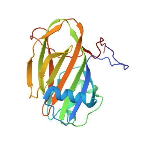
Structure of a 1.5-MDa adhesin that binds its Antarctic bacterium to diatoms and ice.

Guo, S.Stevens, C.A.Vance, T.D.R.Olijve, L.L.C.Graham, L.A.Campbell, R.L.Yazdi, S.R.Escobedo, C.Bar-Dolev, M.Yashunsky, V.Braslavsky, I.Langelaan, D.N.Smith, S.P.Allingham, J.S.Voets, I.K.Davies, P.L.

(2017) Sci Adv 3: e1701440-e1701440

  • DOI: https://doi.org/10.1126/sciadv.1701440
  • Primary Citation of Related Structures:  
    5IRB, 5IX9, 5J6Y, 5JUH, 5K8G

  • PubMed Abstract: 

    Bacterial adhesins are modular cell-surface proteins that mediate adherence to other cells, surfaces, and ligands. The Antarctic bacterium Marinomonas primoryensis uses a 1.5-MDa adhesin comprising over 130 domains to position it on ice at the top of the water column for better access to oxygen and nutrients. We have reconstructed this 0.6-μm-long adhesin using a "dissect and build" structural biology approach and have established complementary roles for its five distinct regions. Domains in region I (RI) tether the adhesin to the type I secretion machinery in the periplasm of the bacterium and pass it through the outer membrane. RII comprises ~120 identical immunoglobulin-like β-sandwich domains that rigidify on binding Ca 2+ to project the adhesion regions RIII and RIV into the medium. RIII contains ligand-binding domains that join diatoms and bacteria together in a mixed-species community on the underside of sea ice where incident light is maximal. RIV is the ice-binding domain, and the terminal RV domain contains several "repeats-in-toxin" motifs and a noncleavable signal sequence that target proteins for export via the type I secretion system. Similar structural architecture is present in the adhesins of many pathogenic bacteria and provides a guide to finding and blocking binding domains to weaken infectivity.


  • Organizational Affiliation

    Protein Function Discovery Group and Department of Biomedical and Molecular Sciences, Queen's University, Kingston, Ontario K7L 3N6, Canada.


Macromolecules
Find similar proteins by:  (by identity cutoff)  |  3D Structure
Entity ID: 1
MoleculeChains Sequence LengthOrganismDetailsImage
Antifreeze protein188Marinomonas primoryensisMutation(s): 0 
UniProt
Find proteins for A1YIY3 (Marinomonas primoryensis)
Explore A1YIY3 
Go to UniProtKB:  A1YIY3
Entity Groups  
Sequence Clusters30% Identity50% Identity70% Identity90% Identity95% Identity100% Identity
UniProt GroupA1YIY3
Sequence Annotations
Expand
  • Reference Sequence
Small Molecules
Ligands 3 Unique
IDChains Name / Formula / InChI Key2D Diagram3D Interactions
GLC
Query on GLC

Download Ideal Coordinates CCD File 
J [auth A]alpha-D-glucopyranose
C6 H12 O6
WQZGKKKJIJFFOK-DVKNGEFBSA-N
BGC
Query on BGC

Download Ideal Coordinates CCD File 
K [auth A]beta-D-glucopyranose
C6 H12 O6
WQZGKKKJIJFFOK-VFUOTHLCSA-N
CA
Query on CA

Download Ideal Coordinates CCD File 
B [auth A]
C [auth A]
D [auth A]
E [auth A]
F [auth A]
CALCIUM ION
Ca
BHPQYMZQTOCNFJ-UHFFFAOYSA-N
Experimental Data & Validation

Experimental Data

  • Method: X-RAY DIFFRACTION
  • Resolution: 1.03 Å
  • R-Value Free:  0.164 (Depositor), 0.160 (DCC) 
  • R-Value Work:  0.155 (Depositor), 0.160 (DCC) 
  • R-Value Observed: 0.156 (Depositor) 
Space Group: P 21 21 21
Unit Cell:
Length ( Å )Angle ( ˚ )
a = 45.22α = 90
b = 50.55β = 90
c = 79.42γ = 90
Software Package:
Software NamePurpose
PHENIXrefinement
XDSdata reduction
Aimlessdata scaling
PHASERphasing

Structure Validation

View Full Validation Report


Ligand Structure Quality Assessment 

Created with Raphaël 2.3.0Worse 01 BetterLigand structure goodness of fit to experimental dataBest fitted GLCClick on this verticalbar to view detailsBest fitted BGCClick on this verticalbar to view details

Entry History 

Deposition Data

  • Released Date: 2017-06-07 
  • Deposition Author(s): Guo, S.

Revision History  (Full details and data files)

  • Version 1.0: 2017-06-07
    Type: Initial release
  • Version 1.1: 2017-09-06
    Changes: Database references
  • Version 2.0: 2020-07-29
    Type: Remediation
    Reason: Carbohydrate remediation
    Changes: Atomic model, Data collection, Derived calculations, Refinement description, Structure summary
  • Version 2.1: 2024-03-06
    Changes: Data collection, Database references, Structure summary