5K8F | pdb_00005k8f

Crystal structure of Acetyl-CoA Synthetase in complex with ATP and Acetyl-AMP from Cryptococcus neoformans H99


Experimental Data Snapshot

  • Method: X-RAY DIFFRACTION
  • Resolution: 2.45 Å
  • R-Value Free: 
    0.224 (Depositor), 0.220 (DCC) 
  • R-Value Work: 
    0.188 (Depositor), 0.190 (DCC) 
  • R-Value Observed: 
    0.189 (Depositor) 

Starting Model: experimental
View more details

wwPDB Validation   3D Report Full Report


Ligand Structure Quality Assessment 

Created with Raphaël 2.3.0Worse 01 BetterLigand structure goodness of fit to experimental dataBest fitted ATPClick on this verticalbar to view detailsBest fitted 6R9Click on this verticalbar to view details

This is version 2.1 of the entry. See complete history


Literature

Crystal structure of Acetyl-CoA Synthetase in complex with ATP and Acetyl-AMP from Cryptococcus neoformans H99

Seattle Structural Genomics Center for Infectious Disease (SSGCID)Fox III, D.Delker, S.L.Potts, K.T.Numa, M.M.Edwards, T.E.Lorimer, D.D.Mutz, M.W.Krysan, D.J.

To be published.

Macromolecules
Find similar proteins by:  (by identity cutoff)  |  3D Structure
Entity ID: 1
MoleculeChains Sequence LengthOrganismDetailsImage
Acetyl-coenzyme A synthetase
A, B, C
694Cryptococcus neoformans var. grubii H99Mutation(s): 0 
Gene Names: CNAG_00797
EC: 6.2.1.1
UniProt
Find proteins for J9VFT1 (Cryptococcus neoformans var. grubii serotype A (strain H99 / ATCC 208821 / CBS 10515 / FGSC 9487))
Explore J9VFT1 
Go to UniProtKB:  J9VFT1
Entity Groups  
Sequence Clusters30% Identity50% Identity70% Identity90% Identity95% Identity100% Identity
UniProt GroupJ9VFT1
Sequence Annotations
Expand
  • Reference Sequence
Small Molecules
Ligands 5 Unique
IDChains Name / Formula / InChI Key2D Diagram3D Interactions
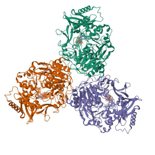
ATP (Subject of Investigation/LOI)
Query on ATP

Download Ideal Coordinates CCD File 
D [auth A],
K [auth B],
Q [auth C]
ADENOSINE-5'-TRIPHOSPHATE
C10 H16 N5 O13 P3
ZKHQWZAMYRWXGA-KQYNXXCUSA-N
6R9 (Subject of Investigation/LOI)
Query on 6R9

Download Ideal Coordinates CCD File 
E [auth A],
L [auth B],
R [auth C]
[[(2~{R},3~{S},4~{R},5~{R})-5-(6-aminopurin-9-yl)-3,4-bis(oxidanyl)oxolan-2-yl]methoxy-oxidanyl-phosphoryl] ethanoate
C12 H16 N5 O8 P
UBPVOHPZRZIJHM-WOUKDFQISA-N
PO4
Query on PO4

Download Ideal Coordinates CCD File 
W [auth C]PHOSPHATE ION
O4 P
NBIIXXVUZAFLBC-UHFFFAOYSA-K
EDO
Query on EDO

Download Ideal Coordinates CCD File 
G [auth A]
H [auth A]
I [auth A]
J [auth A]
N [auth B]
1,2-ETHANEDIOL
C2 H6 O2
LYCAIKOWRPUZTN-UHFFFAOYSA-N
MG
Query on MG

Download Ideal Coordinates CCD File 
F [auth A],
M [auth B],
S [auth C],
V [auth C]
MAGNESIUM ION
Mg
JLVVSXFLKOJNIY-UHFFFAOYSA-N
Experimental Data & Validation

Experimental Data

  • Method: X-RAY DIFFRACTION
  • Resolution: 2.45 Å
  • R-Value Free:  0.224 (Depositor), 0.220 (DCC) 
  • R-Value Work:  0.188 (Depositor), 0.190 (DCC) 
  • R-Value Observed: 0.189 (Depositor) 
Space Group: P 1
Unit Cell:
Length ( Å )Angle ( ˚ )
a = 71.37α = 110.06
b = 83.92β = 105.24
c = 101.57γ = 87.65
Software Package:
Software NamePurpose
PHENIXrefinement
XDSdata reduction
XSCALEdata scaling
PHASERphasing

Structure Validation

View Full Validation Report


Ligand Structure Quality Assessment 

Created with Raphaël 2.3.0Worse 01 BetterLigand structure goodness of fit to experimental dataBest fitted ATPClick on this verticalbar to view detailsBest fitted 6R9Click on this verticalbar to view details

Entry History 

Revision History  (Full details and data files)

  • Version 1.0: 2016-07-13
    Type: Initial release
  • Version 2.0: 2021-04-28
    Type: Coordinate replacement
    Reason: Ligand identity
    Changes: Advisory, Atomic model, Author supporting evidence, Data collection, Database references, Derived calculations, Refinement description, Source and taxonomy, Structure summary
  • Version 2.1: 2023-09-27
    Changes: Data collection, Database references, Refinement description