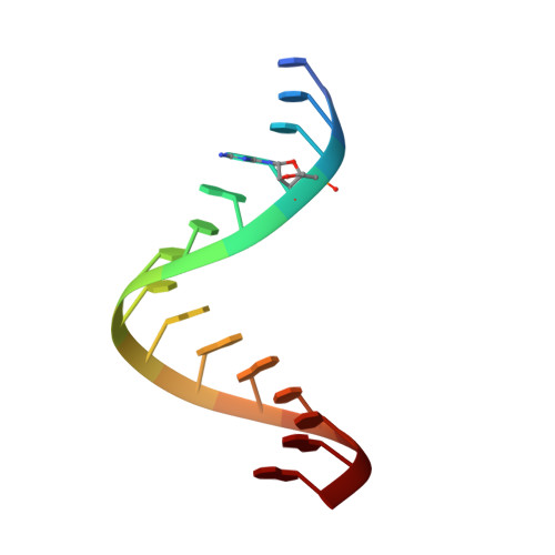
Unusual Base-Pairing Interactions in Monomer-Template Complexes.
Zhang, W., Tam, C.P., Wang, J., Szostak, J.W.(2016) ACS Cent Sci 2: 916-926
- PubMed: 28058281
- DOI: https://doi.org/10.1021/acscentsci.6b00278
- Primary Citation of Related Structures:
5DHB, 5DHC, 5HBW, 5HBX, 5HBY, 5KRG, 5L00 - PubMed Abstract:
Many high-resolution crystal structures have contributed to our understanding of the reaction pathway for catalysis by DNA and RNA polymerases, but the structural basis of nonenzymatic template-directed RNA replication has not been studied in comparable detail. Here we present crystallographic studies of the binding of ribonucleotide monomers to RNA primer-template complexes, with the goal of improving our understanding of the mechanism of nonenzymatic RNA copying, and of catalysis by polymerases. To explore how activated ribonucleotides recognize and bind to RNA templates, we synthesized an unreactive phosphonate-linked pyrazole analogue of guanosine 5'-phosphoro-2-methylimidazolide (2-MeImpG), a highly activated nucleotide that has been used extensively to study nonenzymatic primer extension. We cocrystallized this analogue with structurally rigidified RNA primer-template complexes carrying single or multiple monomer binding sites, and obtained high-resolution X-ray structures of these complexes. In addition to Watson-Crick base pairing, we repeatedly observed noncanonical guanine:cytidine base pairs in our crystal structures. In most structures, the phosphate and leaving group moieties of the monomers were highly disordered, while in others the distance from O3' of the primer to the phosphorus of the incoming monomer was too great to allow for reaction. We suggest that these effects significantly influence the rate and fidelity of nonenzymatic RNA replication, and that even primitive ribozyme polymerases could enhance RNA replication by enforcing Watson-Crick base pairing between monomers and primer-template complexes, and by bringing the reactive functional groups into closer proximity.
Organizational Affiliation:
Howard Hughes Medical Institute, Department of Molecular Biology and Center for Computational and Integrative Biology, Massachusetts General Hospital, 185 Cambridge Street, Boston, Massachusetts 02114, United States; Department of Genetics, Harvard Medical School, 77 Avenue Louis Pasteur, Boston, Massachusetts 02115, United States.


