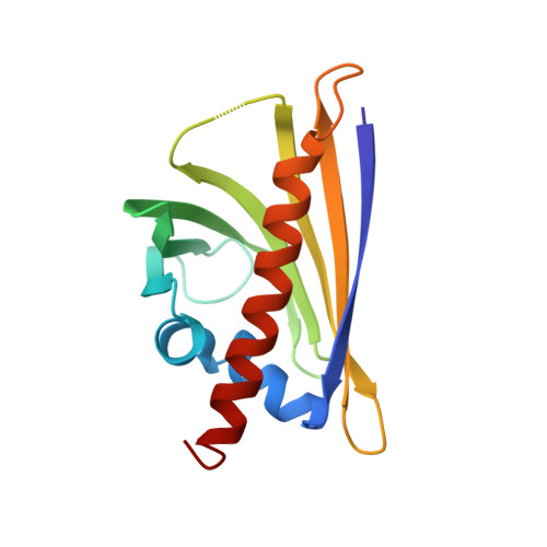
5MXW

Crystal structure of yellow lupin LLPR-10.2B protein in complex with melatonin and trans-zeatin.


Experimental Data Snapshot

  • Method: X-RAY DIFFRACTION
  • Resolution: 1.57 Å
  • R-Value Free: 0.206 
  • R-Value Work: 0.185 
  • R-Value Observed: 0.186 

Starting Model: experimental
View more details

wwPDB Validation   3D Report Full Report


Ligand Structure Quality Assessment 


This is version 1.4 of the entry. See complete history


Literature

PR-10 proteins as potential mediators of melatonin-cytokinin cross-talk in plants: crystallographic studies of LlPR-10.2B isoform from yellow lupine.

Sliwiak, J.Sikorski, M.Jaskolski, M.

(2018) FEBS J 285: 1907-1922

  • DOI: https://doi.org/10.1111/febs.14455
  • Primary Citation of Related Structures:  
    5MXB, 5MXW

  • PubMed Abstract: 

    LlPR-10.2B, a Pathogenesis-related class 10 (PR-10) protein from yellow lupine (Lupinus luteus) was crystallized in complex with melatonin, an emerging important plant regulator and antioxidant. The structure reveals two molecules of melatonin bound in the internal cavity of the protein, plus a very well-defined electron density near the cavity entrance, corresponding to an unknown ligand molecule comprised of two flat rings, which is most likely a product of melatonin transformation. In a separate LlPR-10.2B co-crystallization experiment with an equimolar mixture of melatonin and trans-zeatin, which is a cytokinin phytohormone well recognized as a PR-10-binding partner, a quaternary 1 : 1 : 1 : 1 complex was formed, in which one of the melatonin-binding sites has been substituted with trans-zeatin, whereas the binding of melatonin at the second binding site and binding of the unknown ligand are undisturbed. This unusual complex, when compared with the previously described PR-10/trans-zeatin complexes and with the emerging structural information about melatonin binding by PR-10 proteins, provides intriguing insights into the role of PR-10 proteins in phytohormone regulation in plants, especially with the involvement of melatonin, and implicates the PR-10 proteins as low-affinity melatonin binders under the conditions of elevated melatonin concentration. Atomic coordinates and processed structure factors corresponding to the final models of the LlPR-10.2B/melatonin and LlPR-10.2B/melatonin + trans-zeatin complexes have been deposited with the Protein Data Bank (PDB) under the accession codes 5MXB and 5MXW. The corresponding raw X-ray diffraction images have been deposited in the RepOD Repository at the Interdisciplinary Centre for Mathematical and Computational Modelling (ICM) of the University of Warsaw, Poland, and are available for download with the following Digital Object Identifiers (DOI): https://doi.org/10.18150/repod.9923638 and https://doi.org/10.18150/repod.6621013.


  • Organizational Affiliation

    Center for Biocrystallographic Research, Institute of Bioorganic Chemistry, Polish Academy of Sciences, Poznan, Poland.


Macromolecules
Find similar proteins by:  (by identity cutoff)  |  3D Structure
Entity ID: 1
MoleculeChains Sequence LengthOrganismDetailsImage
Class 10 plant pathogenesis-related protein156Lupinus luteusMutation(s): 0 
Gene Names: pr10.2bYpr10.2b
EC: 3.1.27
UniProt
Find proteins for Q9LLQ2 (Lupinus luteus)
Explore Q9LLQ2 
Go to UniProtKB:  Q9LLQ2
Entity Groups  
Sequence Clusters30% Identity50% Identity70% Identity90% Identity95% Identity100% Identity
UniProt GroupQ9LLQ2
Sequence Annotations
Expand
  • Reference Sequence
Small Molecules
Ligands 4 Unique
IDChains Name / Formula / InChI Key2D Diagram3D Interactions
ML1
Query on ML1

Download Ideal Coordinates CCD File 
S [auth A]N-[2-(5-methoxy-1H-indol-3-yl)ethyl]acetamide
C13 H16 N2 O2
DRLFMBDRBRZALE-UHFFFAOYSA-N
ZEA
Query on ZEA

Download Ideal Coordinates CCD File 
T [auth A](2E)-2-methyl-4-(9H-purin-6-ylamino)but-2-en-1-ol
C10 H13 N5 O
UZKQTCBAMSWPJD-FARCUNLSSA-N
NA
Query on NA

Download Ideal Coordinates CCD File 
R [auth A]SODIUM ION
Na
FKNQFGJONOIPTF-UHFFFAOYSA-N
UNL
Query on UNL

Download Ideal Coordinates CCD File 
B [auth A]
C [auth A]
D [auth A]
E [auth A]
F [auth A]
B [auth A],
C [auth A],
D [auth A],
E [auth A],
F [auth A],
G [auth A],
H [auth A],
I [auth A],
J [auth A],
K [auth A],
L [auth A],
M [auth A],
N [auth A],
O [auth A],
P [auth A],
Q [auth A]
Unknown ligand
FKNQFGJONOIPTF-UHFFFAOYSA-N
Experimental Data & Validation

Experimental Data

Unit Cell:
Length ( Å )Angle ( ˚ )
a = 74.4α = 90
b = 74.4β = 90
c = 67.31γ = 120
Software Package:
Software NamePurpose
REFMACrefinement
XSCALEdata scaling
PDB_EXTRACTdata extraction
XDSdata reduction
PHASERphasing

Structure Validation

View Full Validation Report


Ligand Structure Quality Assessment 


Entry History & Funding Information

Deposition Data


Funding OrganizationLocationGrant Number
European UnionFP7/2007-2013
BioStruct-X283570

Revision History  (Full details and data files)

  • Version 1.0: 2018-04-18
    Type: Initial release
  • Version 1.1: 2018-06-20
    Changes: Data collection, Database references
  • Version 1.2: 2018-08-01
    Changes: Data collection, Derived calculations, Other
  • Version 1.3: 2018-08-08
    Changes: Data collection, Database references
  • Version 1.4: 2024-01-17
    Changes: Data collection, Database references, Refinement description