5N6G

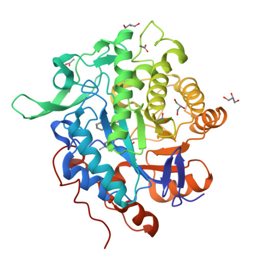
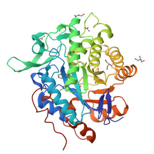
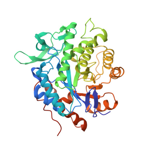
NerA from Agrobacterium radiobacter in complex with 2-phenylacrylic acid


Experimental Data Snapshot

  • Method: X-RAY DIFFRACTION
  • Resolution: 1.58 Å
  • R-Value Free: 0.164 
  • R-Value Work: 0.140 
  • R-Value Observed: 0.142 

Starting Model: experimental
View more details

wwPDB Validation   3D Report Full Report


Ligand Structure Quality Assessment 


This is version 1.3 of the entry. See complete history


Literature

Structural insights into the ene-reductase synthesis of profens.

Waller, J.Toogood, H.S.Karuppiah, V.Rattray, N.J.W.Mansell, D.J.Leys, D.Gardiner, J.M.Fryszkowska, A.Ahmed, S.T.Bandichhor, R.Reddy, G.P.Scrutton, N.S.

(2017) Org Biomol Chem 15: 4440-4448

  • DOI: https://doi.org/10.1039/c7ob00163k
  • Primary Citation of Related Structures:  
    5N6G, 5N6Q

  • PubMed Abstract: 

    Reduction of double bonds of α,β-unsaturated carboxylic acids and esters by ene-reductases remains challenging and it typically requires activation by a second electron-withdrawing moiety, such as a halide or second carboxylate group. We showed that profen precursors, 2-arylpropenoic acids and their esters, were efficiently reduced by Old Yellow Enzymes (OYEs). The XenA and GYE enzymes showed activity towards acids, while a wider range of enzymes were active towards the equivalent methyl esters. Comparative co-crystal structural analysis of profen-bound OYEs highlighted key interactions important in determining substrate binding in a catalytically active conformation. The general utility of ene reductases for the synthesis of (R)-profens was established and this work will now drive future mutagenesis studies to screen for the production of pharmaceutically-active (S)-profens.


  • Organizational Affiliation

    Manchester Institute of Biotechnology, University of Manchester, 131 Princess Street, Manchester M1 7DN, UK. nigel.scrutton@manchester.ac.uk.


Macromolecules
Find similar proteins by:  (by identity cutoff)  |  3D Structure
Entity ID: 1
MoleculeChains Sequence LengthOrganismDetailsImage
GTN Reductase379Agrobacterium tumefaciensMutation(s): 0 
Gene Names: nerA
UniProt
Find proteins for O31246 (Rhizobium radiobacter)
Explore O31246 
Go to UniProtKB:  O31246
Entity Groups  
Sequence Clusters30% Identity50% Identity70% Identity90% Identity95% Identity100% Identity
UniProt GroupO31246
Sequence Annotations
Expand
  • Reference Sequence
Small Molecules
Ligands 4 Unique
IDChains Name / Formula / InChI Key2D Diagram3D Interactions
FMN
Query on FMN

Download Ideal Coordinates CCD File 
B [auth A]FLAVIN MONONUCLEOTIDE
C17 H21 N4 O9 P
FVTCRASFADXXNN-SCRDCRAPSA-N
8OZ
Query on 8OZ

Download Ideal Coordinates CCD File 
C [auth A]2-Phenylacrylic acid
C9 H8 O2
ONPJWQSDZCGSQM-UHFFFAOYSA-N
GOL
Query on GOL

Download Ideal Coordinates CCD File 
D [auth A],
E [auth A],
F [auth A]
GLYCEROL
C3 H8 O3
PEDCQBHIVMGVHV-UHFFFAOYSA-N
ACT
Query on ACT

Download Ideal Coordinates CCD File 
G [auth A],
H [auth A],
I [auth A],
J [auth A]
ACETATE ION
C2 H3 O2
QTBSBXVTEAMEQO-UHFFFAOYSA-M
Experimental Data & Validation

Experimental Data

  • Method: X-RAY DIFFRACTION
  • Resolution: 1.58 Å
  • R-Value Free: 0.164 
  • R-Value Work: 0.140 
  • R-Value Observed: 0.142 
  • Space Group: P 21 21 21
Unit Cell:
Length ( Å )Angle ( ˚ )
a = 60.09α = 90
b = 69.16β = 90
c = 91.89γ = 90
Software Package:
Software NamePurpose
PHENIXrefinement
XDSdata reduction
XSCALEdata scaling
PHASERphasing
PHENIXmodel building

Structure Validation

View Full Validation Report


Ligand Structure Quality Assessment 


Entry History & Funding Information

Deposition Data


Funding OrganizationLocationGrant Number
Biotechnology and Biological Sciences Research CouncilUnited KingdomBB/I015779/1

Revision History  (Full details and data files)

  • Version 1.0: 2017-05-17
    Type: Initial release
  • Version 1.1: 2017-05-31
    Changes: Database references
  • Version 1.2: 2017-08-30
    Changes: Author supporting evidence
  • Version 1.3: 2024-01-17
    Changes: Data collection, Database references, Refinement description