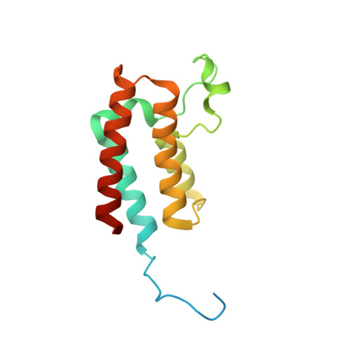
A multi-crystal method for extracting obscured crystallographic states from conventionally uninterpretable electron density.
Pearce, N.M., Krojer, T., Bradley, A.R., Collins, P., Nowak, R.P., Talon, R., Marsden, B.D., Kelm, S., Shi, J., Deane, C.M., von Delft, F.(2017) Nat Commun 8: 15123-15123
- PubMed: 28436492
- DOI: https://doi.org/10.1038/ncomms15123
- Primary Citation of Related Structures:
5PB7, 5PB8, 5PB9, 5PBA, 5PBB, 5PBC, 5PBD, 5PBE, 5PBF, 5PBG, 5PBH, 5PBI, 5PBJ, 5PBK, 5PBL, 5PBM, 5PBN, 5PBO, 5PBP, 5PBQ, 5PBR, 5PBS, 5PBT, 5PBU, 5PBV, 5PBW, 5PBX, 5PBY, 5PBZ, 5PC0, 5PC1, 5PC2, 5PC3, 5PC4, 5PC5, 5PC6, 5PC7, 5PC8, 5PC9, 5PCA, 5PCB, 5PCC, 5PCD, 5PCE, 5PCF, 5PCG, 5PCH, 5PCI, 5PCJ, 5PCK - PubMed Abstract:
In macromolecular crystallography, the rigorous detection of changed states (for example, ligand binding) is difficult unless signal is strong. Ambiguous ('weak' or 'noisy') density is experimentally common, since molecular states are generally only fractionally present in the crystal. Existing methodologies focus on generating maximally accurate maps whereby minor states become discernible; in practice, such map interpretation is disappointingly subjective, time-consuming and methodologically unsound. Here we report the PanDDA method, which automatically reveals clear electron density for the changed state-even from inaccurate maps-by subtracting a proportion of the confounding 'ground state'; changed states are objectively identified from statistical analysis of density distributions. The method is completely general, implying new best practice for all changed-state studies, including the routine collection of multiple ground-state crystals. More generally, these results demonstrate: the incompleteness of atomic models; that single data sets contain insufficient information to model them fully; and that accuracy requires further map-deconvolution approaches.
Organizational Affiliation:
Structural Genomics Consortium, Nuffield Department of Medicine, University of Oxford, Roosevelt Drive, Oxford OX3 7DQ, UK.


