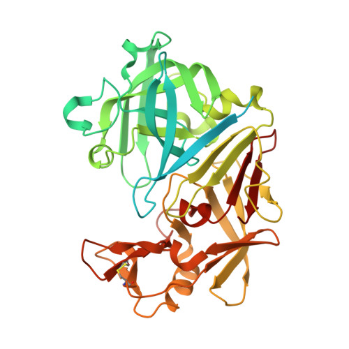
Frag4Lead: growing crystallographic fragment hits by catalog using fragment-guided template docking.
Metz, A., Wollenhaupt, J., Glockner, S., Messini, N., Huber, S., Barthel, T., Merabet, A., Gerber, H.D., Heine, A., Klebe, G., Weiss, M.S.(2021) Acta Crystallogr D Struct Biol 77: 1168-1182
- PubMed: 34473087
- DOI: https://doi.org/10.1107/S2059798321008196
- Primary Citation of Related Structures:
5SAK, 5SAL, 5SAM, 5SAN, 5SAO, 5SAP, 5SAQ, 5SAR, 5SAS, 5SAT - PubMed Abstract:
In recent years, crystallographic fragment screening has matured into an almost routine experiment at several modern synchrotron sites. The hits of the screening experiment, i.e. small molecules or fragments binding to the target protein, are revealed along with their 3D structural information. Therefore, they can serve as useful starting points for further structure-based hit-to-lead development. However, the progression of fragment hits to tool compounds or even leads is often hampered by a lack of chemical feasibility. As an attractive alternative, compound analogs that embed the fragment hit structurally may be obtained from commercial catalogs. Here, a workflow is reported based on filtering and assessing such potential follow-up compounds by template docking. This means that the crystallographic binding pose was integrated into the docking calculations as a central starting parameter. Subsequently, the candidates are scored on their interactions within the binding pocket. In an initial proof-of-concept study using five starting fragments known to bind to the aspartic protease endothiapepsin, 28 follow-up compounds were selected using the designed workflow and their binding was assessed by crystallography. Ten of these compounds bound to the active site and five of them showed significantly increased affinity in isothermal titration calorimetry of up to single-digit micromolar affinity. Taken together, this strategy is capable of efficiently evolving the initial fragment hits without major synthesis efforts and with full control by X-ray crystallography.
Organizational Affiliation:
Department of Pharmaceutical Chemistry, Philipps-University Marburg, Marbacher Weg 6, D-35032 Marburg, Germany.


