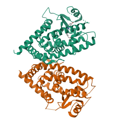
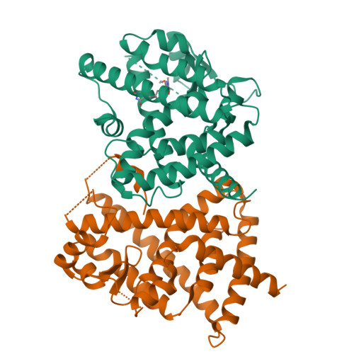
5U5L

X-ray Crystal Structure of the PPARgamma Ligand Binding Domain in Complex with Rivoglitazone


Experimental Data Snapshot

  • Method: X-RAY DIFFRACTION
  • Resolution: 2.55 Å
  • R-Value Free: 0.238 
  • R-Value Work: 0.188 
  • R-Value Observed: 0.191 

Starting Model: experimental
View more details

wwPDB Validation   3D Report Full Report


Ligand Structure Quality Assessment 


This is version 1.1 of the entry. See complete history


Literature

X-ray crystal structure of rivoglitazone bound to PPAR gamma and PPAR subtype selectivity of TZDs.

Rajapaksha, H.Bhatia, H.Wegener, K.Petrovsky, N.Bruning, J.B.

(2017) Biochim Biophys Acta 1861: 1981-1991

  • DOI: https://doi.org/10.1016/j.bbagen.2017.05.008
  • Primary Citation of Related Structures:  
    5U5L

  • PubMed Abstract: 

    Thiazolidinedione (TZD) compounds targeting the nuclear receptor peroxisome proliferator-activated receptor gamma (PPARγ) demonstrate unique benefits for the treatment of insulin resistance and type II diabetes. TZDs include rosiglitazone, pioglitazone and rivoglitazone, with the latter being the most potent. The TZDs are only marginally selective for the therapeutic target PPARγ as they also activate PPARα and PPARδ homologues to varying degrees, causing off-target effects. While crystal structures for TZD compounds in complex with PPARγ are available, minimal structural information is available for TZDs bound to PPARα and PPARδ. This paucity of structural information has hampered the determination of precise structural mechanisms involved in TZD selectivity between PPARs. To help address these questions molecular dynamic simulations were performed of rosiglitazone, pioglitazone and rivoglitazone in complex with PPARα, PPARδ, and PPARγ in order to better understand the mechanisms of PPAR selectivity. The simulations revealed that TZD interactions with residues Tyr314 and Phe318 of PPARα and residues Phe291 and Thr253 of PPARδ as well as the omega loop, are key determinants of TZD receptor selectivity. Notably, in this study, we solve the first X-ray crystal structure of rivoglitazone bound to any PPAR. Rivoglitazone forms a unique hydrogen bond network with the residues of the PPARγ co-activator binding surface (known as AF2) and makes more extensive contacts with helix 3 and the β-sheet as compared to model TZD compounds such as rosiglitazone.


  • Organizational Affiliation

    Department of Diabetes and Endocrinology, School of Medicine, Flinders University, Adelaide, South Australia 5042, Australia.


Macromolecules
Find similar proteins by:  (by identity cutoff)  |  3D Structure
Entity ID: 1
MoleculeChains Sequence LengthOrganismDetailsImage
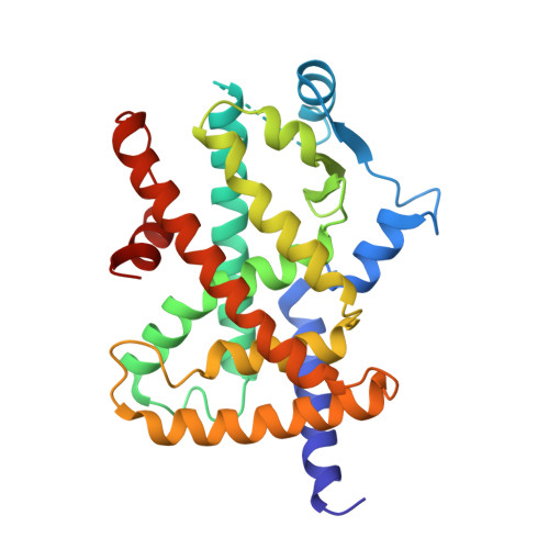
Peroxisome proliferator-activated receptor gamma
A, B
273Homo sapiensMutation(s): 0 
Gene Names: PPARGNR1C3
UniProt & NIH Common Fund Data Resources
Find proteins for P37231 (Homo sapiens)
Explore P37231 
Go to UniProtKB:  P37231
PHAROS:  P37231
GTEx:  ENSG00000132170 
Entity Groups  
Sequence Clusters30% Identity50% Identity70% Identity90% Identity95% Identity100% Identity
UniProt GroupP37231
Sequence Annotations
Expand
  • Reference Sequence
Small Molecules
Ligands 1 Unique
IDChains Name / Formula / InChI Key2D Diagram3D Interactions
7VA
Query on 7VA

Download Ideal Coordinates CCD File 
C [auth A](5S)-5-({4-[(6-methoxy-1-methyl-1H-benzimidazol-2-yl)methoxy]phenyl}methyl)-1,3-thiazolidine-2,4-dione
C20 H19 N3 O4 S
XMSXOLDPMGMWTH-UHFFFAOYSA-N
Experimental Data & Validation

Experimental Data

  • Method: X-RAY DIFFRACTION
  • Resolution: 2.55 Å
  • R-Value Free: 0.238 
  • R-Value Work: 0.188 
  • R-Value Observed: 0.191 
  • Space Group: C 1 2 1
Unit Cell:
Length ( Å )Angle ( ˚ )
a = 93.394α = 90
b = 62.097β = 102.22
c = 118.47γ = 90
Software Package:
Software NamePurpose
Aimlessdata scaling
PHASERphasing
PHENIXrefinement
PDB_EXTRACTdata extraction
XDSdata reduction

Structure Validation

View Full Validation Report


Ligand Structure Quality Assessment 


Entry History 

Deposition Data

Revision History  (Full details and data files)

  • Version 1.0: 2017-08-23
    Type: Initial release
  • Version 1.1: 2023-10-04
    Changes: Data collection, Database references, Refinement description