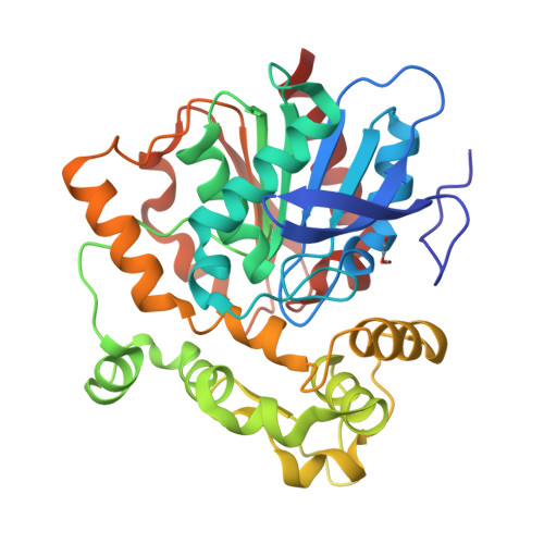
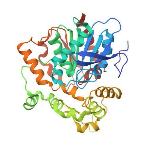
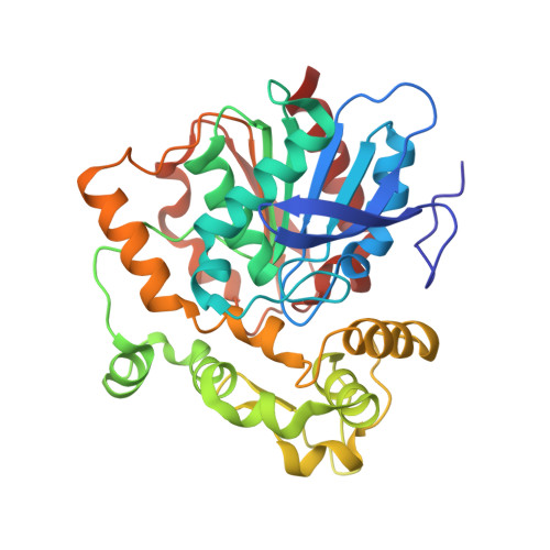
5URO

Structure of a soluble epoxide hydrolase identified in Trichoderma reesei


Experimental Data Snapshot

  • Method: X-RAY DIFFRACTION
  • Resolution: 1.70 Å
  • R-Value Free: 0.211 
  • R-Value Work: 0.183 
  • R-Value Observed: 0.184 

Starting Model: experimental
View more details

wwPDB Validation   3D Report Full Report


This is version 1.4 of the entry. See complete history


Literature

Structure of a soluble epoxide hydrolase identified in Trichoderma reesei.

Wilson, C.De Oliveira, G.S.Adriani, P.P.Chambergo, F.S.Dias, M.V.B.

(2017) Biochim Biophys Acta 1865: 1039-1045

  • DOI: https://doi.org/10.1016/j.bbapap.2017.05.004
  • Primary Citation of Related Structures:  
    5URO

  • PubMed Abstract: 

    Epoxide hydrolases (EHs) are enzymes that have high biotechnological interest for the fine and transformation industry. Several of these enzymes have enantioselectivity, which allows their application in the separation of enantiomeric mixtures of epoxide substrates. Although two different families of EHs have been described, those that have the α/β-hidrolase fold are the most explored for biotechnological purpose. These enzymes are functionally very well studied, but only few members have three-dimensional structures characterised. Recently, a new EH from the filamentous fungi Trichoderma reseei (TrEH) has been discovered and functionally studied. This enzyme does not have high homology to any other EH structure and have an enatiopreference for (S)-(-) isomers. Herein we described the crystallographic structure of TrEH at 1.7Å resolution, which reveals features of its tertiary structure and active site. TrEH has a similar fold to the other soluble epoxide hydrolases and has the two characteristic hydrolase and cap domains. The enzyme is predominantly monomeric in solution and has also been crystallised as a monomer in the asymmetric unit. Although the catalytic residues are conserved, several other residues of the catalytic groove are not, and might be involved in the specificity for substrates and in the enantioselectivy of this enzyme. In addition, the determination of the crystallographic structure of TrEH might contribute to the rational site direct mutagenesis to generate an even more stable enzyme with higher efficiency to be used in biotechnological purposes.


  • Organizational Affiliation

    Department of Microbiology, Institute of Biomedical Science, University of São Paulo, São Paulo, Brazil; Programa de Pós Graduação em Microbiologia, IBILCE-UNESP, São José do Rio Preto, São Paulo, Brazil.


Macromolecules
Find similar proteins by:  (by identity cutoff)  |  3D Structure
Entity ID: 1
MoleculeChains Sequence LengthOrganismDetailsImage
Predicted protein336Trichoderma reesei QM6aMutation(s): 0 
Gene Names: TRIREDRAFT_53220
UniProt
Find proteins for G0R7E2 (Hypocrea jecorina (strain QM6a))
Explore G0R7E2 
Go to UniProtKB:  G0R7E2
Entity Groups  
Sequence Clusters30% Identity50% Identity70% Identity90% Identity95% Identity100% Identity
UniProt GroupG0R7E2
Sequence Annotations
Expand
  • Reference Sequence
Small Molecules
Ligands 1 Unique
IDChains Name / Formula / InChI Key2D Diagram3D Interactions
8LD
Query on 8LD

Download Ideal Coordinates CCD File 
B [auth A]2,2'-oxydi(ethyn-1-ol)
C4 H2 O3
QFHUYNIAGZXZMU-UHFFFAOYSA-N
Experimental Data & Validation

Experimental Data

  • Method: X-RAY DIFFRACTION
  • Resolution: 1.70 Å
  • R-Value Free: 0.211 
  • R-Value Work: 0.183 
  • R-Value Observed: 0.184 
  • Space Group: P 2 21 21
Unit Cell:
Length ( Å )Angle ( ˚ )
a = 51.427α = 90
b = 78.281β = 90
c = 87.372γ = 90
Software Package:
Software NamePurpose
Aimlessdata scaling
PHASERphasing
PDB_EXTRACTdata extraction
XDSdata reduction
PHENIXrefinement

Structure Validation

View Full Validation Report


Entry History & Funding Information

Deposition Data


Funding OrganizationLocationGrant Number
Sao Paulo Research Foundation (FAPESP)Brazil--

Revision History  (Full details and data files)

  • Version 1.0: 2017-05-31
    Type: Initial release
  • Version 1.1: 2017-06-21
    Changes: Database references
  • Version 1.2: 2019-04-17
    Changes: Author supporting evidence, Data collection
  • Version 1.3: 2020-01-01
    Changes: Author supporting evidence
  • Version 1.4: 2023-10-04
    Changes: Data collection, Database references, Refinement description