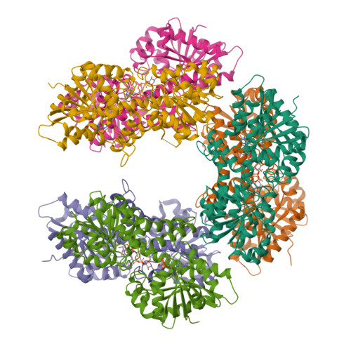
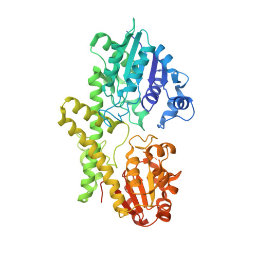
5VR8

Human UDP-Glucose Dehydrogenase with UDP-Xylose Bound to the Co-enzyme Site


Experimental Data Snapshot

  • Method: X-RAY DIFFRACTION
  • Resolution: 2.00 Å
  • R-Value Free: 0.190 
  • R-Value Work: 0.156 
  • R-Value Observed: 0.158 

wwPDB Validation   3D Report Full Report


Ligand Structure Quality Assessment 


This is version 2.7 of the entry. See complete history


Literature

The entropic force generated by intrinsically disordered segments tunes protein function.

Keul, N.D.Oruganty, K.Schaper Bergman, E.T.Beattie, N.R.McDonald, W.E.Kadirvelraj, R.Gross, M.L.Phillips, R.S.Harvey, S.C.Wood, Z.A.

(2018) Nature 563: 584-588

  • DOI: https://doi.org/10.1038/s41586-018-0699-5
  • Primary Citation of Related Structures:  
    5VR8, 5W4X

  • PubMed Abstract: 

    Protein structures are dynamic and can explore a large conformational landscape 1,2 . Only some of these structural substates are important for protein function (such as ligand binding, catalysis and regulation) 3-5 . How evolution shapes the structural ensemble to optimize a specific function is poorly understood 3,4 . One of the constraints on the evolution of proteins is the stability of the folded 'native' state. Despite this, 44% of the human proteome contains intrinsically disordered peptide segments greater than 30 residues in length 6 , the majority of which have no known function 7-9 . Here we show that the entropic force produced by an intrinsically disordered carboxy terminus (ID-tail) shifts the conformational ensemble of human UDP-α-D-glucose-6-dehydrogenase (UGDH) towards a substate with a high affinity for an allosteric inhibitor. The function of the ID-tail does not depend on its sequence or chemical composition. Instead, the affinity enhancement can be accurately predicted based on the length of the intrinsically disordered segment, and is consistent with the entropic force generated by an unstructured peptide attached to the protein surface 10-13 . Our data show that the unfolded state of the ID-tail rectifies the dynamics and structure of UGDH to favour inhibitor binding. Because this entropic rectifier does not have any sequence or structural constraints, it is an easily acquired adaptation. This model implies that evolution selects for disordered segments to tune the energy landscape of proteins, which may explain the persistence of intrinsic disorder in the proteome.


  • Organizational Affiliation

    Department of Biochemistry and Molecular Biology, University of Georgia, Athens, GA, USA.


Macromolecules
Find similar proteins by:  (by identity cutoff)  |  3D Structure
Entity ID: 1
MoleculeChains Sequence LengthOrganismDetailsImage
UDP-glucose 6-dehydrogenase
A, B, C, D, E
A, B, C, D, E, F
495Homo sapiensMutation(s): 0 
Gene Names: UGDH
EC: 1.1.1.22
UniProt & NIH Common Fund Data Resources
Find proteins for O60701 (Homo sapiens)
Explore O60701 
Go to UniProtKB:  O60701
PHAROS:  O60701
GTEx:  ENSG00000109814 
Entity Groups  
Sequence Clusters30% Identity50% Identity70% Identity90% Identity95% Identity100% Identity
UniProt GroupO60701
Sequence Annotations
Expand
  • Reference Sequence
Small Molecules
Ligands 2 Unique
IDChains Name / Formula / InChI Key2D Diagram3D Interactions
UDX
Query on UDX

Download Ideal Coordinates CCD File 
G [auth A]
H [auth A]
I [auth B]
J [auth B]
L [auth C]
G [auth A],
H [auth A],
I [auth B],
J [auth B],
L [auth C],
M [auth D],
N [auth D],
O [auth E],
P [auth E],
R [auth F]
URIDINE-5'-DIPHOSPHATE-XYLOPYRANOSE
C14 H22 N2 O16 P2
DQQDLYVHOTZLOR-OCIMBMBZSA-N
ADP
Query on ADP

Download Ideal Coordinates CCD File 
K [auth C],
Q [auth F]
ADENOSINE-5'-DIPHOSPHATE
C10 H15 N5 O10 P2
XTWYTFMLZFPYCI-KQYNXXCUSA-N
Experimental Data & Validation

Experimental Data

  • Method: X-RAY DIFFRACTION
  • Resolution: 2.00 Å
  • R-Value Free: 0.190 
  • R-Value Work: 0.156 
  • R-Value Observed: 0.158 
  • Space Group: P 1 21 1
Unit Cell:
Length ( Å )Angle ( ˚ )
a = 89.08α = 90
b = 196.49β = 111.91
c = 111.26γ = 90
Software Package:
Software NamePurpose
PHENIXrefinement
XDSdata reduction
XDSdata scaling
PHASERphasing

Structure Validation

View Full Validation Report


Ligand Structure Quality Assessment 


Entry History 

Deposition Data

Revision History  (Full details and data files)

  • Version 1.0: 2017-07-19
    Type: Initial release
  • Version 2.0: 2018-03-21
    Changes: Advisory, Database references, Polymer sequence, Source and taxonomy, Structure summary
  • Version 2.1: 2018-03-28
    Changes: Data collection, Database references
  • Version 2.2: 2018-11-07
    Changes: Data collection, Database references
  • Version 2.3: 2018-11-14
    Changes: Data collection, Database references
  • Version 2.4: 2018-11-28
    Changes: Data collection, Database references
  • Version 2.5: 2018-12-05
    Changes: Data collection, Database references
  • Version 2.6: 2023-04-26
    Changes: Database references, Derived calculations
  • Version 2.7: 2024-05-22
    Changes: Data collection