5WFV

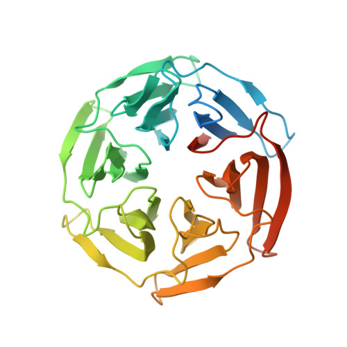
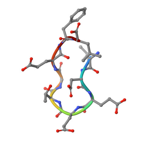
Kelch domain of human Keap1 bound to Nrf2 ETGE peptide


Experimental Data Snapshot

  • Method: X-RAY DIFFRACTION
  • Resolution: 1.91 Å
  • R-Value Free: 0.227 
  • R-Value Work: 0.200 
  • R-Value Observed: 0.200 

Starting Model: experimental
View more details

wwPDB Validation   3D Report Full Report


This is version 1.3 of the entry. See complete history


Literature

Interaction Energetics and Druggability of the Protein-Protein Interaction between Kelch-like ECH-Associated Protein 1 (KEAP1) and Nuclear Factor Erythroid 2 Like 2 (Nrf2).

Zhong, M.Lynch, A.Muellers, S.N.Jehle, S.Luo, L.Hall, D.R.Iwase, R.Carolan, J.P.Egbert, M.Wakefield, A.Streu, K.Harvey, C.M.Ortet, P.C.Kozakov, D.Vajda, S.Allen, K.N.Whitty, A.

(2020) Biochemistry 59: 563-581

  • DOI: https://doi.org/10.1021/acs.biochem.9b00943
  • Primary Citation of Related Structures:  
    5WFL, 5WFV, 5WG1, 5WHL, 5WHO, 5WIY

  • PubMed Abstract: 

    Development of small molecule inhibitors of protein-protein interactions (PPIs) is hampered by our poor understanding of the druggability of PPI target sites. Here, we describe the combined application of alanine-scanning mutagenesis, fragment screening, and FTMap computational hot spot mapping to evaluate the energetics and druggability of the highly charged PPI interface between Kelch-like ECH-associated protein 1 (KEAP1) and nuclear factor erythroid 2 like 2 (Nrf2), an important drug target. FTMap identifies four binding energy hot spots at the active site. Only two of these are exploited by Nrf2, which alanine scanning of both proteins shows to bind primarily through E79 and E82 interacting with KEAP1 residues S363, R380, R415, R483, and S508. We identify fragment hits and obtain X-ray complex structures for three fragments via crystal soaking using a new crystal form of KEAP1. Combining these results provides a comprehensive and quantitative picture of the origins of binding energy at the interface. Our findings additionally reveal non-native interactions that might be exploited in the design of uncharged synthetic ligands to occupy the same site on KEAP1 that has evolved to bind the highly charged DEETGE binding loop of Nrf2. These include π-stacking with KEAP1 Y525 and interactions at an FTMap-identified hot spot deep in the binding site. Finally, we discuss how the complementary information provided by alanine-scanning mutagenesis, fragment screening, and computational hot spot mapping can be integrated to more comprehensively evaluate PPI druggability.


  • Organizational Affiliation

    Acpharis, Inc. , 160 North Mill Street , Holliston , Massachusetts 01746 , United States.


Macromolecules
Find similar proteins by:  (by identity cutoff)  |  3D Structure
Entity ID: 1
MoleculeChains Sequence LengthOrganismDetailsImage
Kelch-like ECH-associated protein 1
A, B
336Homo sapiensMutation(s): 2 
Gene Names: KEAP1INRF2KIAA0132KLHL19
UniProt & NIH Common Fund Data Resources
Find proteins for Q14145 (Homo sapiens)
Explore Q14145 
Go to UniProtKB:  Q14145
PHAROS:  Q14145
GTEx:  ENSG00000079999 
Entity Groups  
Sequence Clusters30% Identity50% Identity70% Identity90% Identity95% Identity100% Identity
UniProt GroupQ14145
Sequence Annotations
Expand
  • Reference Sequence

Find similar proteins by:  Sequence   |   3D Structure  

Entity ID: 2
MoleculeChains Sequence LengthOrganismDetailsImage
Nrf2 ETGE peptideC [auth P]9Homo sapiensMutation(s): 0 
UniProt & NIH Common Fund Data Resources
Find proteins for Q16236 (Homo sapiens)
Explore Q16236 
Go to UniProtKB:  Q16236
PHAROS:  Q16236
GTEx:  ENSG00000116044 
Entity Groups  
UniProt GroupQ16236
Sequence Annotations
Expand
  • Reference Sequence
Small Molecules
Ligands 1 Unique
IDChains Name / Formula / InChI Key2D Diagram3D Interactions
SO4
Query on SO4

Download Ideal Coordinates CCD File 
D [auth B]SULFATE ION
O4 S
QAOWNCQODCNURD-UHFFFAOYSA-L
Experimental Data & Validation

Experimental Data

  • Method: X-RAY DIFFRACTION
  • Resolution: 1.91 Å
  • R-Value Free: 0.227 
  • R-Value Work: 0.200 
  • R-Value Observed: 0.200 
  • Space Group: C 1 2 1
Unit Cell:
Length ( Å )Angle ( ˚ )
a = 162.919α = 90
b = 69.253β = 118.5
c = 79.228γ = 90
Software Package:
Software NamePurpose
PHENIXrefinement
XDSdata reduction
XDSdata scaling
PHASERphasing

Structure Validation

View Full Validation Report


Entry History & Funding Information

Deposition Data


Funding OrganizationLocationGrant Number
National Institutes of Health/National Institute of General Medical Sciences (NIH/NIGMS)United StatesR35-GM118078

Revision History  (Full details and data files)

  • Version 1.0: 2018-09-19
    Type: Initial release
  • Version 1.1: 2020-01-01
    Changes: Author supporting evidence
  • Version 1.2: 2020-04-01
    Changes: Database references
  • Version 1.3: 2023-10-04
    Changes: Data collection, Database references, Refinement description