5WGK | pdb_00005wgk

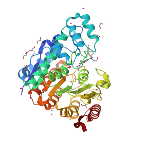
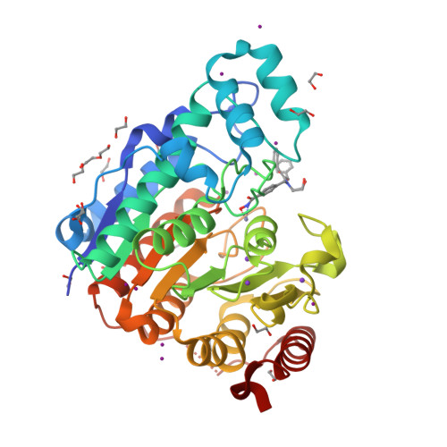
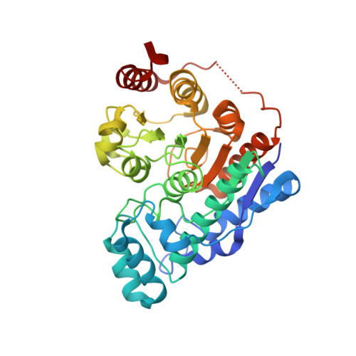
Crystal structure of Danio rerio histone deacetylase 6 catalytic domain 2 in complex with HPB


Experimental Data Snapshot

  • Method: X-RAY DIFFRACTION
  • Resolution: 1.82 Å
  • R-Value Free: 
    0.173 (Depositor), 0.180 (DCC) 
  • R-Value Work: 
    0.149 (Depositor), 0.150 (DCC) 
  • R-Value Observed: 
    0.150 (Depositor) 

Starting Model: experimental
View more details

wwPDB Validation   3D Report Full Report


Ligand Structure Quality Assessment 

Created with Raphaël 2.3.0Worse 01 BetterLigand structure goodness of fit to experimental dataBest fitted AGJClick on this verticalbar to view details

This is version 1.4 of the entry. See complete history


Literature

Unusual zinc-binding mode of HDAC6-selective hydroxamate inhibitors.

Porter, N.J.Mahendran, A.Breslow, R.Christianson, D.W.

(2017) Proc Natl Acad Sci U S A 114: 13459-13464

  • DOI: https://doi.org/10.1073/pnas.1718823114
  • Primary Citation of Related Structures:  
    5WGI, 5WGK, 5WGL, 5WGM

  • PubMed Abstract: 

    Histone deacetylases (HDACs) regulate myriad cellular processes by catalyzing the hydrolysis of acetyl-l-lysine residues in histone and nonhistone proteins. The Zn 2+ -dependent class IIb enzyme HDAC6 regulates microtubule function by deacetylating α-tubulin, which suppresses microtubule dynamics and leads to cell cycle arrest and apoptosis. Accordingly, HDAC6 is a target for the development of selective inhibitors that might be useful in new therapeutic approaches for the treatment of cancer, neurodegenerative diseases, and other disorders. Here, we present high-resolution structures of catalytic domain 2 from Danio rerio HDAC6 (henceforth simply "HDAC6") complexed with compounds that selectively inhibit HDAC6 while maintaining nanomolar inhibitory potency: N -hydroxy-4-[( N (2-hydroxyethyl)-2-phenylacetamido)methyl)-benzamide)] (HPB), ACY-1215 (Ricolinostat), and ACY-1083. These structures reveal that an unusual monodentate Zn 2+ coordination mode is exploited by sterically bulky HDAC6-selective phenylhydroxamate inhibitors. We additionally report the ultrahigh-resolution structure of the HDAC6-trichostatin A complex, which reveals two Zn 2+ -binding conformers for the inhibitor: a major conformer (70%) with canonical bidentate hydroxamate-Zn 2+ coordination geometry and a minor conformer (30%) with monodentate hydroxamate-Zn 2+ coordination geometry, reflecting a free energy difference of only 0.5 kcal/mol. The minor conformer is not visible in lower resolution structure determinations. Structural comparisons of HDAC6-inhibitor complexes with class I HDACs suggest active site features that contribute to the isozyme selectivity observed in biochemical assays.


  • Organizational Affiliation

    Roy and Diana Vagelos Laboratories, Department of Chemistry, University of Pennsylvania, Philadelphia, PA 19104-6323.


Macromolecules
Find similar proteins by:  (by identity cutoff)  |  3D Structure
Entity ID: 1
MoleculeChains Sequence LengthOrganismDetailsImage
Hdac6 protein364Danio rerioMutation(s): 0 
Gene Names: hdac6
UniProt
Find proteins for F8W4B7 (Danio rerio)
Explore F8W4B7 
Go to UniProtKB:  F8W4B7
Entity Groups  
Sequence Clusters30% Identity50% Identity70% Identity90% Identity95% Identity100% Identity
UniProt GroupF8W4B7
Sequence Annotations
Expand
  • Reference Sequence
Small Molecules
Ligands 6 Unique
IDChains Name / Formula / InChI Key2D Diagram3D Interactions
AGJ
Query on AGJ

Download Ideal Coordinates CCD File 
Q [auth A]N-hydroxy-4-{[(2-hydroxyethyl)(phenylacetyl)amino]methyl}benzamide
C18 H20 N2 O4
GTFAUKGKYICUDS-UHFFFAOYSA-N
IOD
Query on IOD

Download Ideal Coordinates CCD File 
B [auth A]
C [auth A]
D [auth A]
E [auth A]
F [auth A]
IODIDE ION
I
XMBWDFGMSWQBCA-UHFFFAOYSA-M
PEG
Query on PEG

Download Ideal Coordinates CCD File 
FA [auth A]DI(HYDROXYETHYL)ETHER
C4 H10 O3
MTHSVFCYNBDYFN-UHFFFAOYSA-N
ZN
Query on ZN

Download Ideal Coordinates CCD File 
M [auth A]ZINC ION
Zn
PTFCDOFLOPIGGS-UHFFFAOYSA-N
EDO
Query on EDO

Download Ideal Coordinates CCD File 
AA [auth A]
BA [auth A]
CA [auth A]
DA [auth A]
EA [auth A]
1,2-ETHANEDIOL
C2 H6 O2
LYCAIKOWRPUZTN-UHFFFAOYSA-N
K
Query on K

Download Ideal Coordinates CCD File 
N [auth A],
O [auth A],
P [auth A]
POTASSIUM ION
K
NPYPAHLBTDXSSS-UHFFFAOYSA-N
Experimental Data & Validation

Experimental Data

  • Method: X-RAY DIFFRACTION
  • Resolution: 1.82 Å
  • R-Value Free:  0.173 (Depositor), 0.180 (DCC) 
  • R-Value Work:  0.149 (Depositor), 0.150 (DCC) 
  • R-Value Observed: 0.150 (Depositor) 
Space Group: P 2 21 21
Unit Cell:
Length ( Å )Angle ( ˚ )
a = 51.67α = 90
b = 83.97β = 90
c = 94.57γ = 90
Software Package:
Software NamePurpose
PHENIXrefinement
iMOSFLMdata reduction
Aimlessdata scaling
PHASERphasing

Structure Validation

View Full Validation Report


Ligand Structure Quality Assessment 

Created with Raphaël 2.3.0Worse 01 BetterLigand structure goodness of fit to experimental dataBest fitted AGJClick on this verticalbar to view details

Entry History & Funding Information

Deposition Data


Funding OrganizationLocationGrant Number
National Institutes of Health/National Institute of General Medical Sciences (NIH/NIGMS)United StatesGM49758

Revision History  (Full details and data files)

  • Version 1.0: 2017-12-06
    Type: Initial release
  • Version 1.1: 2017-12-20
    Changes: Database references
  • Version 1.2: 2018-01-03
    Changes: Database references
  • Version 1.3: 2020-01-01
    Changes: Author supporting evidence
  • Version 1.4: 2023-10-04
    Changes: Data collection, Database references, Derived calculations, Refinement description