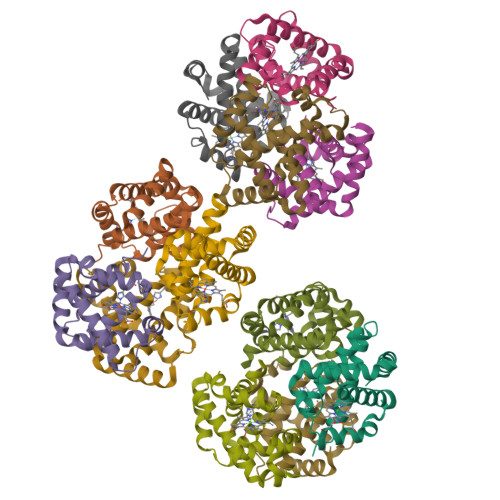
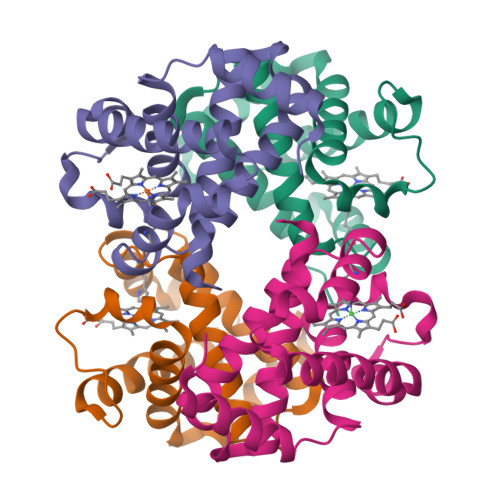
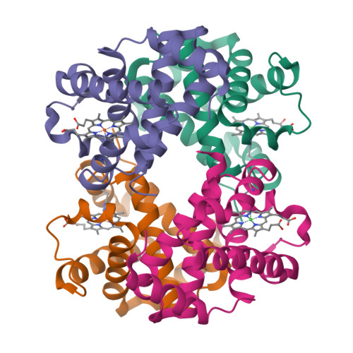
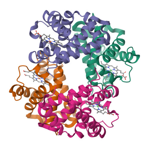
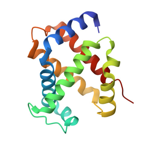
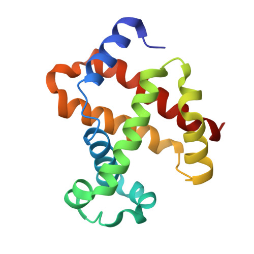
5X2U

Direct Observation of Conformational Population Shifts in Hemoglobin: Crystal Structure of Half-Liganded Hemoglobin after Adding 80 mM phosphate pH 6.7.


Experimental Data Snapshot

  • Method: X-RAY DIFFRACTION
  • Resolution: 2.53 Å
  • R-Value Free: 0.274 
  • R-Value Work: 0.231 
  • R-Value Observed: 0.233 

wwPDB Validation   3D Report Full Report


Ligand Structure Quality Assessment 


This is version 1.2 of the entry. See complete history


Literature

Direct observation of conformational population shifts in crystalline human hemoglobin.

Shibayama, N.Ohki, M.Tame, J.R.H.Park, S.Y.

(2017) J Biol Chem 292: 18258-18269

  • DOI: https://doi.org/10.1074/jbc.M117.781146
  • Primary Citation of Related Structures:  
    5X2R, 5X2S, 5X2T, 5X2U

  • PubMed Abstract: 

    Although X-ray crystallography is the most commonly used technique for studying the molecular structure of proteins, it is not generally able to monitor the dynamic changes or global domain motions that often underlie allostery. These motions often prevent crystal growth or reduce crystal order. We have recently discovered a crystal form of human hemoglobin that contains three protein molecules allowed to express a full range of quaternary structures, whereas maintaining strong X-ray diffraction. Here we use this crystal form to investigate the effects of two allosteric effectors, phosphate and bezafibrate, by tracking the structures and functions of the three hemoglobin molecules following the addition of each effector. The X-ray analysis shows that the addition of either phosphate or bezafibrate not only induces conformational changes in a direction from a relaxed-state to a tense-state, but also within relaxed-state populations. The microspectrophotometric O 2 equilibrium measurements on the crystals demonstrate that the binding of each effector energetically stabilizes the lowest affinity conformer more strongly than the intermediate affinity one, thereby reducing the O 2 affinity of tense-state populations, and that the addition of bezafibrate causes an ∼5-fold decrease in the O 2 affinity of relaxed-state populations. These results show that the allosteric pathway of hemoglobin involves shifts of populations rather than a unidirectional conversion of one quaternary structure to another, and that minor conformers of hemoglobin may have a disproportionate effect on the overall O 2 affinity.


  • Organizational Affiliation

    From the Department of Physiology, Division of Biophysics, Jichi Medical University, 3311-1 Yakushiji, Shimotsuke, Tochigi 329-0498 and shibayam@jichi.ac.jp.


Macromolecules
Find similar proteins by:  (by identity cutoff)  |  3D Structure
Entity ID: 1
MoleculeChains Sequence LengthOrganismDetailsImage
Hemoglobin subunit alpha
A, C, E, G, I
A, C, E, G, I, K
141Homo sapiensMutation(s): 0 
UniProt & NIH Common Fund Data Resources
Find proteins for P69905 (Homo sapiens)
Explore P69905 
Go to UniProtKB:  P69905
PHAROS:  P69905
Entity Groups  
Sequence Clusters30% Identity50% Identity70% Identity90% Identity95% Identity100% Identity
UniProt GroupP69905
Sequence Annotations
Expand
  • Reference Sequence
Find similar proteins by:  (by identity cutoff)  |  3D Structure
Entity ID: 2
MoleculeChains Sequence LengthOrganismDetailsImage
Hemoglobin subunit beta
B, D, F, H, J
B, D, F, H, J, L
146Homo sapiensMutation(s): 0 
UniProt & NIH Common Fund Data Resources
Find proteins for P68871 (Homo sapiens)
Explore P68871 
Go to UniProtKB:  P68871
PHAROS:  P68871
GTEx:  ENSG00000244734 
Entity Groups  
Sequence Clusters30% Identity50% Identity70% Identity90% Identity95% Identity100% Identity
UniProt GroupP68871
Sequence Annotations
Expand
  • Reference Sequence
Small Molecules
Ligands 2 Unique
IDChains Name / Formula / InChI Key2D Diagram3D Interactions
HNI
Query on HNI

Download Ideal Coordinates CCD File 
M [auth A]
P [auth D]
Q [auth E]
T [auth H]
U [auth I]
M [auth A],
P [auth D],
Q [auth E],
T [auth H],
U [auth I],
X [auth L]
PROTOPORPHYRIN IX CONTAINING NI(II)
C34 H32 N4 Ni O4
IJROJBFZULUEEG-RGGAHWMASA-L
HEM
Query on HEM

Download Ideal Coordinates CCD File 
N [auth B]
O [auth C]
R [auth F]
S [auth G]
V [auth J]
N [auth B],
O [auth C],
R [auth F],
S [auth G],
V [auth J],
W [auth K]
PROTOPORPHYRIN IX CONTAINING FE
C34 H32 Fe N4 O4
KABFMIBPWCXCRK-RGGAHWMASA-L
Experimental Data & Validation

Experimental Data

  • Method: X-RAY DIFFRACTION
  • Resolution: 2.53 Å
  • R-Value Free: 0.274 
  • R-Value Work: 0.231 
  • R-Value Observed: 0.233 
  • Space Group: C 1 2 1
Unit Cell:
Length ( Å )Angle ( ˚ )
a = 228.886α = 90
b = 55.812β = 102.39
c = 139.963γ = 90
Software Package:
Software NamePurpose
BUSTERrefinement
HKL-2000data reduction
HKL-2000data scaling

Structure Validation

View Full Validation Report


Ligand Structure Quality Assessment 


Entry History 

Deposition Data

Revision History  (Full details and data files)

  • Version 1.0: 2017-09-27
    Type: Initial release
  • Version 1.1: 2018-02-21
    Changes: Database references
  • Version 1.2: 2024-03-27
    Changes: Data collection, Database references