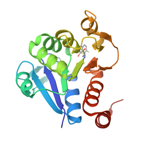
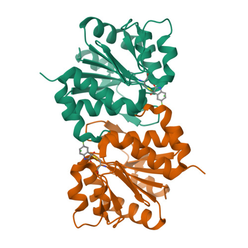
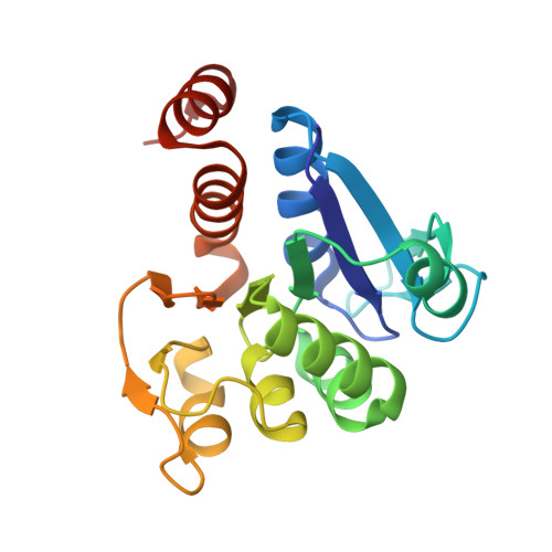
6AFC

DJ-1 with compound 4


Experimental Data Snapshot

  • Method: X-RAY DIFFRACTION
  • Resolution: 1.45 Å
  • R-Value Free: 0.160 
  • R-Value Work: 0.133 
  • R-Value Observed: 0.134 

Starting Model: experimental
View more details

wwPDB Validation   3D Report Full Report


Ligand Structure Quality Assessment 


This is version 1.3 of the entry. See complete history


Literature

Discovery and Optimization of Inhibitors of the Parkinson's Disease Associated Protein DJ-1.

Tashiro, S.Caaveiro, J.M.M.Nakakido, M.Tanabe, A.Nagatoishi, S.Tamura, Y.Matsuda, N.Liu, D.Hoang, Q.Q.Tsumoto, K.

(2018) ACS Chem Biol 13: 2783-2793

  • DOI: https://doi.org/10.1021/acschembio.8b00701
  • Primary Citation of Related Structures:  
    6AF5, 6AF7, 6AF9, 6AFA, 6AFB, 6AFC, 6AFD, 6AFE, 6AFF, 6AFG, 6AFH, 6AFI, 6AFJ, 6AFL

  • PubMed Abstract: 

    DJ-1 is a Parkinson's disease associated protein endowed with enzymatic, redox sensing, regulatory, chaperoning, and neuroprotective activities. Although DJ-1 has been vigorously studied for the past decade and a half, its exact role in the progression of the disease remains uncertain. In addition, little is known about the spatiotemporal regulation of DJ-1, or the biochemical basis explaining its numerous biological functions. Progress has been hampered by the lack of inhibitors with precisely known mechanisms of action. Herein, we have employed biophysical methodologies and X-ray crystallography to identify and to optimize a family of compounds inactivating the critical Cys106 residue of human DJ-1. We demonstrate these compounds are potent inhibitors of various activities of DJ-1 in vitro and in cell-based assays. This study reports a new family of DJ-1 inhibitors with a defined mechanism of action, and contributes toward the understanding of the biological function of DJ-1.


  • Organizational Affiliation

    Department of Bioengineering, Graduate School of Engineering , The University of Tokyo , Tokyo 113-8656 , Japan.


Macromolecules
Find similar proteins by:  (by identity cutoff)  |  3D Structure
Entity ID: 1
MoleculeChains Sequence LengthOrganismDetailsImage
Protein/nucleic acid deglycase DJ-1189Homo sapiensMutation(s): 0 
Gene Names: PARK7
EC: 3.1.2 (PDB Primary Data), 3.5.1 (PDB Primary Data), 3.5.1.124 (PDB Primary Data)
UniProt & NIH Common Fund Data Resources
Find proteins for Q99497 (Homo sapiens)
Explore Q99497 
Go to UniProtKB:  Q99497
PHAROS:  Q99497
GTEx:  ENSG00000116288 
Entity Groups  
Sequence Clusters30% Identity50% Identity70% Identity90% Identity95% Identity100% Identity
UniProt GroupQ99497
Sequence Annotations
Expand
  • Reference Sequence
Experimental Data & Validation

Experimental Data

  • Method: X-RAY DIFFRACTION
  • Resolution: 1.45 Å
  • R-Value Free: 0.160 
  • R-Value Work: 0.133 
  • R-Value Observed: 0.134 
  • Space Group: P 31 2 1
Unit Cell:
Length ( Å )Angle ( ˚ )
a = 75.43α = 90
b = 75.43β = 90
c = 75.89γ = 120
Software Package:
Software NamePurpose
REFMACrefinement
MOSFLMdata reduction
SCALAdata scaling
PHASERphasing

Structure Validation

View Full Validation Report


Ligand Structure Quality Assessment 


Entry History & Funding Information

Deposition Data


Funding OrganizationLocationGrant Number
Ministry of Education, Culture, Sports, Science and Technology (Japan)JapanPlatform for Drug Discovery, Informatics and Structural Life Science
Japan Society for the Promotion of ScienceJapan25249115
Japan Society for the Promotion of ScienceJapan16H02420
Japan Society for the Promotion of ScienceJapan15K06962

Revision History  (Full details and data files)

  • Version 1.0: 2018-08-29
    Type: Initial release
  • Version 1.1: 2018-10-03
    Changes: Data collection, Database references
  • Version 1.2: 2023-11-22
    Changes: Data collection, Database references, Refinement description
  • Version 1.3: 2024-10-16
    Changes: Structure summary