6ATJ

RECOMBINANT HORSERADISH PEROXIDASE C COMPLEX WITH FERULIC ACID


Experimental Data Snapshot

  • Method: X-RAY DIFFRACTION
  • Resolution: 2.00 Å
  • R-Value Free: 0.199 
  • R-Value Work: 0.166 

Starting Model: experimental
View more details

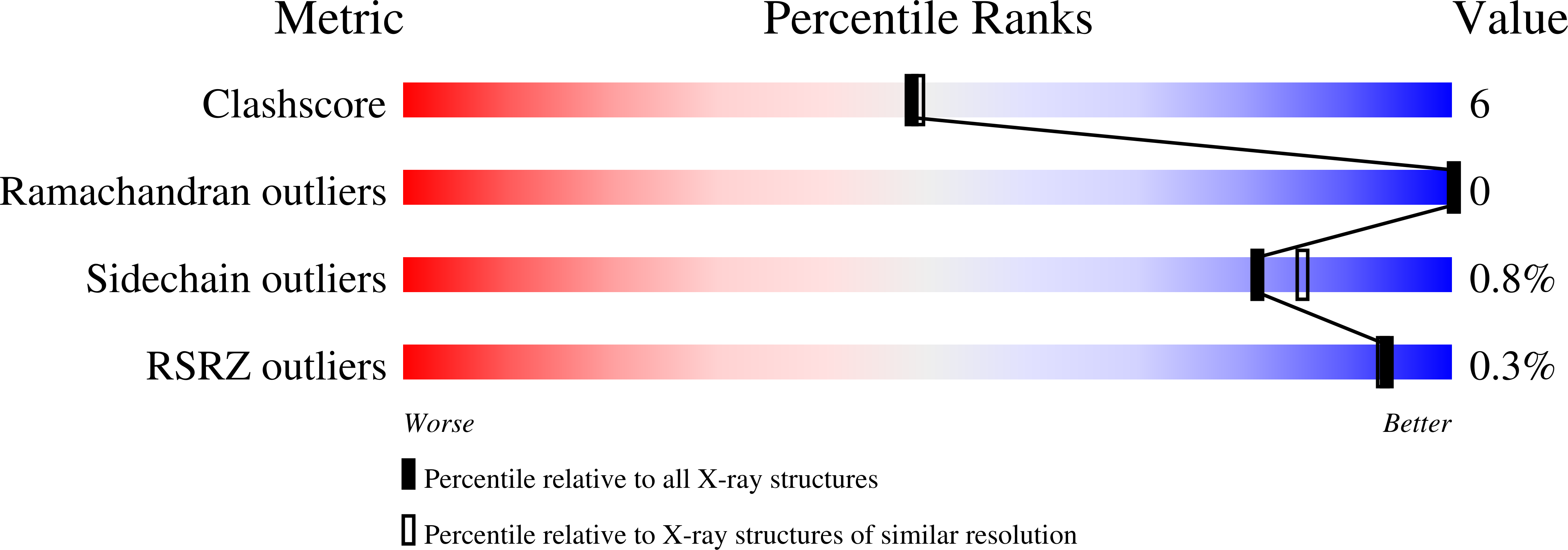
wwPDB Validation   3D Report Full Report


Ligand Structure Quality Assessment 


This is version 1.5 of the entry. See complete history


Literature

The structures of the horseradish peroxidase C-ferulic acid complex and the ternary complex with cyanide suggest how peroxidases oxidize small phenolic substrates.

Henriksen, A.Smith, A.T.Gajhede, M.

(1999) J Biol Chem 274: 35005-35011

  • DOI: https://doi.org/10.1074/jbc.274.49.35005
  • Primary Citation of Related Structures:  
    6ATJ, 7ATJ

  • PubMed Abstract: 

    We have solved the x-ray structures of the binary horseradish peroxidase C-ferulic acid complex and the ternary horseradish peroxidase C-cyanide-ferulic acid complex to 2.0 and 1.45 A, respectively. Ferulic acid is a naturally occurring phenolic compound found in the plant cell wall and is an in vivo substrate for plant peroxidases. The x-ray structures demonstrate the flexibility and dynamic character of the aromatic donor binding site in horseradish peroxidase and emphasize the role of the distal arginine (Arg(38)) in both substrate oxidation and ligand binding. Arg(38) hydrogen bonds to bound cyanide, thereby contributing to the stabilization of the horseradish peroxidase-cyanide complex and suggesting that the distal arginine will be able to contribute with a similar interaction during stabilization of a bound peroxy transition state and subsequent O-O bond cleavage. The catalytic arginine is additionally engaged in an extensive hydrogen bonding network, which also includes the catalytic distal histidine, a water molecule and Pro(139), a proline residue conserved within the plant peroxidase superfamily. Based on the observed hydrogen bonding network and previous spectroscopic and kinetic work, a general mechanism of peroxidase substrate oxidation is proposed.


  • Organizational Affiliation

    Protein Structure Group, Department of Chemistry, University of Copenhagen, Universitetsparken 5, DK-2100 Kobenhavn O, Denmark. anette@jerne.ki.ku.dk


Macromolecules
Find similar proteins by:  (by identity cutoff)  |  3D Structure
Entity ID: 1
MoleculeChains Sequence LengthOrganismDetailsImage
PEROXIDASE C1A308Armoracia rusticanaMutation(s): 0 
EC: 1.11.1.7
UniProt
Find proteins for P00433 (Armoracia rusticana)
Explore P00433 
Go to UniProtKB:  P00433
Entity Groups  
Sequence Clusters30% Identity50% Identity70% Identity90% Identity95% Identity100% Identity
UniProt GroupP00433
Sequence Annotations
Expand
  • Reference Sequence
Experimental Data & Validation

Experimental Data

  • Method: X-RAY DIFFRACTION
  • Resolution: 2.00 Å
  • R-Value Free: 0.199 
  • R-Value Work: 0.166 
  • Space Group: P 21 21 21
Unit Cell:
Length ( Å )Angle ( ˚ )
a = 40.297α = 90
b = 67.272β = 90
c = 117.646γ = 90
Software Package:
Software NamePurpose
DENZOdata reduction
SCALEPACKdata scaling
AMoREphasing
CNSrefinement

Structure Validation

View Full Validation Report


Ligand Structure Quality Assessment 


Entry History 

Deposition Data

Revision History  (Full details and data files)

  • Version 1.0: 2000-01-14
    Type: Initial release
  • Version 1.1: 2007-10-16
    Changes: Version format compliance
  • Version 1.2: 2011-07-13
    Changes: Version format compliance
  • Version 1.3: 2019-11-27
    Changes: Database references
  • Version 1.4: 2023-09-20
    Changes: Data collection, Database references, Derived calculations, Refinement description
  • Version 1.5: 2024-11-20
    Changes: Structure summary