6CWW | pdb_00006cww

Cs H-NOX mutant with unnatural amino acid 4-cyano-L-phenylalanine at site 5


Experimental Data Snapshot

  • Method: X-RAY DIFFRACTION
  • Resolution: 1.85 Å
  • R-Value Free: 
    0.259 (Depositor), 0.260 (DCC) 
  • R-Value Work: 
    0.191 (Depositor), 0.190 (DCC) 
  • R-Value Observed: 
    0.195 (Depositor) 

Starting Model: experimental
View more details

wwPDB Validation   3D Report Full Report


Ligand Structure Quality Assessment 

Created with Raphaël 2.3.0Worse 01 BetterLigand structure goodness of fit to experimental dataBest fitted 211Click on this verticalbar to view details

This is version 1.5 of the entry. See complete history


Literature

Exploring local solvation environments of a heme protein using the spectroscopic reporter 4-cyano-l-phenylalanine.

Kearney, C.Olenginski, L.T.Hirn, T.D.Fowler, G.D.Tariq, D.Brewer, S.H.Phillips-Piro, C.M.

(2018) RSC Adv 8: 13503-13512

  • DOI: https://doi.org/10.1039/c8ra02000k
  • Primary Citation of Related Structures:  
    6CWW

  • PubMed Abstract: 

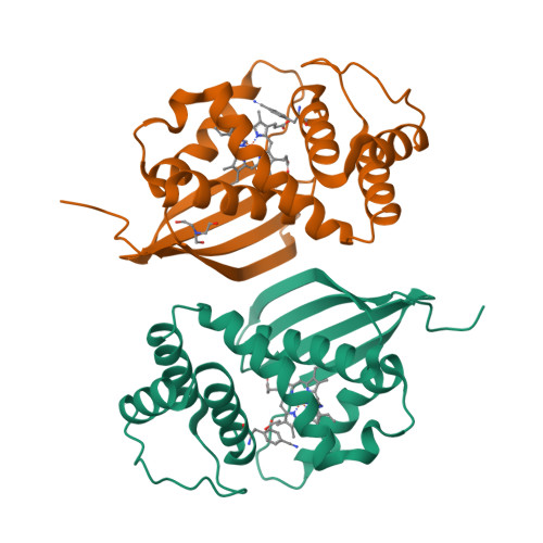
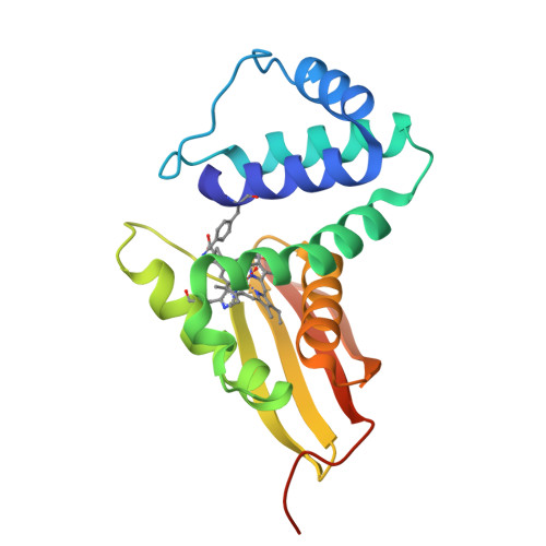
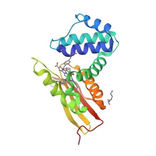
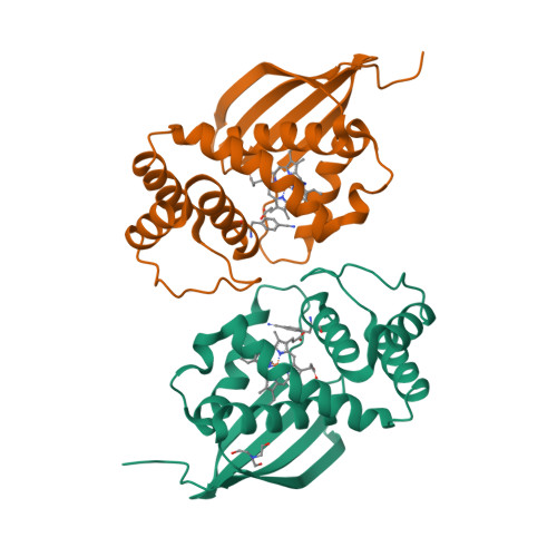
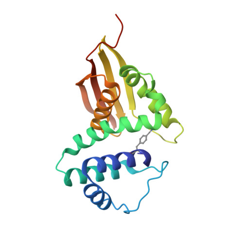
    The vibrational reporter unnatural amino acid (UAA) 4-cyano-l-phenylalanine (pCNF) was genetically incorporated individually at three sites (5, 36, and 78) in the heme protein Caldanaerobacter subterraneus H-NOX to probe local hydration environments. The UAA pCNF was incorporated site-specifically using an engineered, orthogonal tRNA synthetase in E. coli . The ability of all of the pCNF-containing H-NOX proteins to form the ferrous CO, NO, or O 2 ligated and unligated states was confirmed with UV-Vis spectroscopy. The solvation state at each site of the three sites of pCNF incorporation was assessed using temperature-dependent infrared spectroscopy. Specifically, the frequency-temperature line slope (FTLS) method was utilized to show that the nitrile group at site 36 was fully solvated and the nitrile group at site 78 was de-solvated (buried) in the heme pocket. The nitrile group at site 5 was found to be partially solvated suggesting that the nitrile group was involved in moderate strength hydrogen bonds. These results were confirmed by the determination of the X-ray crystal structure of the H-NOX protein construct containing pCNF at site 5.


  • Organizational Affiliation

    Department of Chemistry, Franklin & Marshall College, PO Box 3003, Lancaster, PA 17604-3003, USA. Email: sbrewer@fandm.edu; Email: cpiro@fandm.edu.


Macromolecules
Find similar proteins by:  (by identity cutoff)  |  3D Structure
Entity ID: 1
MoleculeChains Sequence LengthOrganismDetailsImage
Methyl-accepting chemotaxis protein
A, B
194Caldanaerobacter subterraneusMutation(s): 1 
Gene Names: XD49_1849
UniProt
Find proteins for Q8RBX6 (Caldanaerobacter subterraneus subsp. tengcongensis (strain DSM 15242 / JCM 11007 / NBRC 100824 / MB4))
Explore Q8RBX6 
Go to UniProtKB:  Q8RBX6
Entity Groups  
Sequence Clusters30% Identity50% Identity70% Identity90% Identity95% Identity100% Identity
UniProt GroupQ8RBX6
Sequence Annotations
Expand
  • Reference Sequence
Small Molecules
Experimental Data & Validation

Experimental Data

  • Method: X-RAY DIFFRACTION
  • Resolution: 1.85 Å
  • R-Value Free:  0.259 (Depositor), 0.260 (DCC) 
  • R-Value Work:  0.191 (Depositor), 0.190 (DCC) 
  • R-Value Observed: 0.195 (Depositor) 
Space Group: P 1 21 1
Unit Cell:
Length ( Å )Angle ( ˚ )
a = 51.055α = 90
b = 67.129β = 105.33
c = 57.669γ = 90
Software Package:
Software NamePurpose
PHENIXrefinement
HKL-2000data reduction
HKL-2000data scaling
PHASERphasing

Structure Validation

View Full Validation Report


Ligand Structure Quality Assessment 

Created with Raphaël 2.3.0Worse 01 BetterLigand structure goodness of fit to experimental dataBest fitted 211Click on this verticalbar to view details

Entry History & Funding Information

Deposition Data


Funding OrganizationLocationGrant Number
National Institutes of Health/National Institute of General Medical Sciences (NIH/NIGMS)United StatesR15GM121984
National Science Foundation (NSF, United States)United StatesCHE-1053946

Revision History  (Full details and data files)

  • Version 1.0: 2018-05-16
    Type: Initial release
  • Version 1.1: 2018-06-06
    Changes: Data collection, Database references
  • Version 1.2: 2019-11-27
    Changes: Author supporting evidence
  • Version 1.3: 2023-10-04
    Changes: Data collection, Database references, Derived calculations, Refinement description
  • Version 1.4: 2023-11-15
    Changes: Data collection
  • Version 1.5: 2024-10-30
    Changes: Structure summary