6IKA | pdb_00006ika

HIV-1 reverse transcriptase with Q151M/G112S/D113A/Y115F/F116Y/F160L/I159L:DNA:entecavir-triphosphate ternary complex


Experimental Data Snapshot

  • Method: X-RAY DIFFRACTION
  • Resolution: 2.60 Å
  • R-Value Free: 
    0.232 (Depositor), 0.230 (DCC) 
  • R-Value Work: 
    0.186 (Depositor), 0.190 (DCC) 
  • R-Value Observed: 
    0.188 (Depositor) 

Starting Model: experimental
View more details

wwPDB Validation   3D Report Full Report


Ligand Structure Quality Assessment 

Created with Raphaël 2.3.0Worse 01 BetterLigand structure goodness of fit to experimental dataBest fitted ET9Click on this verticalbar to view details

This is version 1.2 of the entry. See complete history


Literature

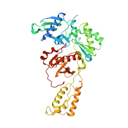
Active-site deformation in the structure of HIV-1 RT with HBV-associated septuple amino acid substitutions rationalizes the differential susceptibility of HIV-1 and HBV against 4'-modified nucleoside RT inhibitors.

Yasutake, Y.Hattori, S.I.Tamura, N.Matsuda, K.Kohgo, S.Maeda, K.Mitsuya, H.

(2019) Biochem Biophys Res Commun 509: 943-948

  • DOI: https://doi.org/10.1016/j.bbrc.2019.01.026
  • Primary Citation of Related Structures:  
    6IK9, 6IKA

  • PubMed Abstract: 

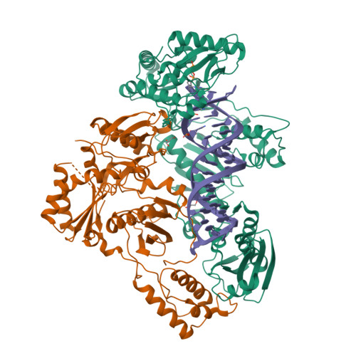
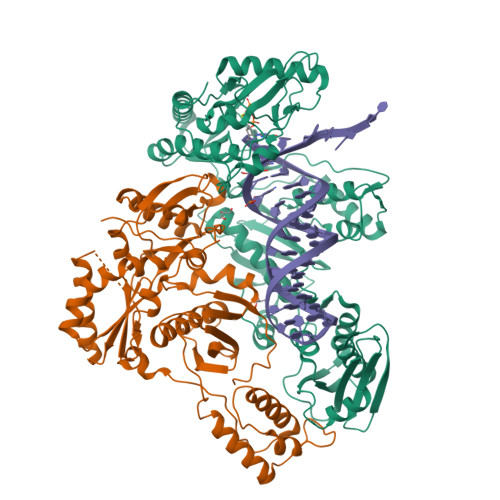
    Nucleoside analogue reverse transcriptase (RT) inhibitors (NRTIs) are major antiviral agents against hepatitis B virus (HBV) and human immunodeficiency virus type-1 (HIV-1). However, the notorious insoluble property of HBV RT has prevented atomic-resolution structural studies and rational anti-HBV drug design. Here, we created HIV-1 RT mutants containing HBV-mimicking sextuple or septuple amino acid substitutions at the nucleoside-binding site (N-site) and verified that these mutants retained the RT activity. The most active RT mutant, HIV-1 RT 7MC , carrying Q151M/G112S/D113A/Y115F/F116Y/F160L/I159L was successfully crystallized, and its three-dimensional structure was determined in complex with DNA:dGTP/entecavir-triphosphate (ETV-TP), a potent anti-HBV guanosine analogue RT inhibitor, at a resolution of 2.43 Å and 2.60 Å, respectively. The structures reveal significant positional rearrangements of the amino acid side-chains at the N-site, elucidating the mechanism underlying the differential susceptibility of HIV-1 and HBV against recently reported 4'-modified NRTIs.


  • Organizational Affiliation

    Bioproduction Research Institute, National Institute of Advanced Industrial Science and Technology (AIST), Sapporo, 062-8517, Japan; Computational Bio Big-Data Open Innovation Laboratory (CBBD-OIL), AIST, Sapporo, 062-8517, Japan. Electronic address: y-yasutake@aist.go.jp.


Macromolecules

Find similar proteins by:  (by identity cutoff)  |  3D Structure
Entity ID: 1
MoleculeChains Sequence LengthOrganismDetailsImage
HIV-1 RT p66 subunitA,
D [auth C]
557Human immunodeficiency virus 1Mutation(s): 9 
UniProt
Find proteins for D3XFN7 (Human immunodeficiency virus type 1)
Explore D3XFN7 
Go to UniProtKB:  D3XFN7
Entity Groups  
Sequence Clusters30% Identity50% Identity70% Identity90% Identity95% Identity100% Identity
UniProt GroupD3XFN7
Sequence Annotations
Expand
  • Reference Sequence
Find similar proteins by:  (by identity cutoff)  |  3D Structure
Entity ID: 2
MoleculeChains Sequence LengthOrganismDetailsImage
HIV-1 RT p51 subunitB,
E [auth D]
444Human immunodeficiency virus 1Mutation(s): 2 
UniProt
Find proteins for P12497 (Human immunodeficiency virus type 1 group M subtype B (isolate NY5))
Explore P12497 
Go to UniProtKB:  P12497
Entity Groups  
Sequence Clusters30% Identity50% Identity70% Identity90% Identity95% Identity100% Identity
UniProt GroupP12497
Sequence Annotations
Expand
  • Reference Sequence
Small Molecules
Ligands 3 Unique
IDChains Name / Formula / InChI Key2D Diagram3D Interactions
ET9
Query on ET9

Download Ideal Coordinates CCD File 
G [auth A],
K [auth C]
[[(1R,3S,5S)-3-(2-azanyl-6-oxidanylidene-3H-purin-9-yl)-2-methylidene-5-oxidanyl-cyclopentyl]methoxy-oxidanyl-phosphory l] phosphono hydrogen phosphate
C12 H18 N5 O12 P3
YMBBDUCQYPKKJK-FXQIFTODSA-N
GOL
Query on GOL

Download Ideal Coordinates CCD File 
H [auth A],
J [auth B],
L [auth C],
N [auth D]
GLYCEROL
C3 H8 O3
PEDCQBHIVMGVHV-UHFFFAOYSA-N
MG
Query on MG

Download Ideal Coordinates CCD File 
I [auth A],
M [auth C]
MAGNESIUM ION
Mg
JLVVSXFLKOJNIY-UHFFFAOYSA-N
Experimental Data & Validation

Experimental Data

  • Method: X-RAY DIFFRACTION
  • Resolution: 2.60 Å
  • R-Value Free:  0.232 (Depositor), 0.230 (DCC) 
  • R-Value Work:  0.186 (Depositor), 0.190 (DCC) 
  • R-Value Observed: 0.188 (Depositor) 
Space Group: H 3
Unit Cell:
Length ( Å )Angle ( ˚ )
a = 285.405α = 90
b = 285.405β = 90
c = 96.346γ = 120
Software Package:
Software NamePurpose
PHENIXrefinement
XDSdata reduction
XDSdata scaling
PHENIXphasing

Structure Validation

View Full Validation Report


Ligand Structure Quality Assessment 

Created with Raphaël 2.3.0Worse 01 BetterLigand structure goodness of fit to experimental dataBest fitted ET9Click on this verticalbar to view details

Entry History & Funding Information

Deposition Data


Funding OrganizationLocationGrant Number
Japan Agency for Medical Research and Development (AMED)JapanJP18fk0310113

Revision History  (Full details and data files)

  • Version 1.0: 2019-01-30
    Type: Initial release
  • Version 1.1: 2019-02-06
    Changes: Data collection, Database references
  • Version 1.2: 2023-11-22
    Changes: Data collection, Database references, Derived calculations, Refinement description, Structure summary