6KHP

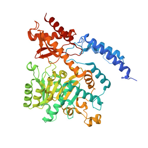
Crystal structure of Oryza sativa TDC with PLP and tryptamine


Experimental Data Snapshot

  • Method: X-RAY DIFFRACTION
  • Resolution: 2.30 Å
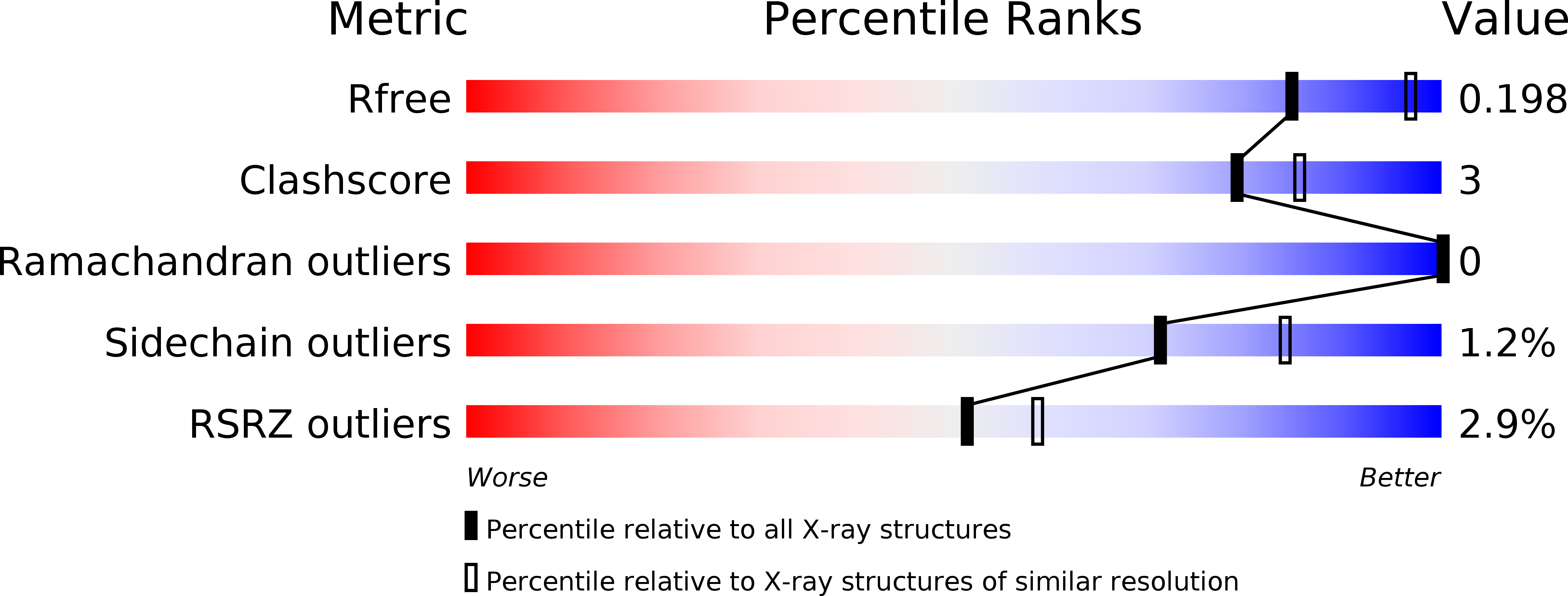
  • R-Value Free: 0.198 
  • R-Value Work: 0.163 
  • R-Value Observed: 0.165 

wwPDB Validation   3D Report Full Report


Ligand Structure Quality Assessment 


This is version 1.0 of the entry. See complete history


Literature

Crystal structure ofOryza sativaTDC reveals the substrate specificity for TDC-mediated melatonin biosynthesis.

Zhou, Y.Liao, L.Liu, X.Liu, B.Chen, X.Guo, Y.Huang, C.Zhao, Y.Zeng, Z.

(2020) J Adv Res 24: 501-511

  • DOI: https://doi.org/10.1016/j.jare.2020.06.004
  • Primary Citation of Related Structures:  
    6KHN, 6KHO, 6KHP

  • PubMed Abstract: 

    Plant tryptophan decarboxylase (TDC) is a type II Pyridoxal-5'-phosphate-dependent decarboxylase (PLP_DC) that could be used as a target to genetically improve crops. However, lack of accurate structural information on plant TDC hampers the understanding of its decarboxylation mechanisms. In the present study, the crystal structures of Oryza sativa TDC ( O sTDC) in its complexes with pyridoxal-5'-phosphate, tryptamine and serotonin were determined. The structures provide detailed interaction information between TDC and its substrates. The Y359 residue from the loop gate is a proton donor and forms a Lewis acid-base pair with serotonin/tryptamine, which is associated with product release. The H214 residue is responsible for PLP binding and proton transfer, and its proper interaction with Y359 is essential for Os TDC enzyme activity. The extra hydrogen bonds formed between the 5-hydroxyl group of serotonin and the backbone carboxyl groups of F104 and P105 explain the discrepancy between the catalytic activity of TDC in tryptophan and in 5-hydroxytryptophan. In addition, an evolutionary analysis revealed that type II PLP_DC originated from glutamic acid decarboxylase, potentially as an adaptive evolution of mechanism in organisms in extreme environments. This study is, to our knowledge, the first to present a detailed analysis of the crystal structure of Os TDC in these complexes. The information regarding the catalytic mechanism described here could facilitate the development of protocols to regulate melatonin levels and thereby contribute to crop improvement efforts to improve food security worldwide.


  • Organizational Affiliation

    National Key Laboratory of Crop Genetic Improvement, Huazhong Agricultural University, Wuhan 430070, China.


Macromolecules
Find similar proteins by:  (by identity cutoff)  |  3D Structure
Entity ID: 1
MoleculeChains Sequence LengthOrganismDetailsImage
Tryptophan decarboxylase 1
A, B
514Oryza sativa Japonica GroupMutation(s): 0 
Gene Names: TDC1Os08g0140300LOC_Os08g04540OJ1368_G08.14OsJ_25993
EC: 4.1.1 (PDB Primary Data), 4.1.1.28 (UniProt)
UniProt
Find proteins for Q6ZJK7 (Oryza sativa subsp. japonica)
Explore Q6ZJK7 
Go to UniProtKB:  Q6ZJK7
Entity Groups  
Sequence Clusters30% Identity50% Identity70% Identity90% Identity95% Identity100% Identity
UniProt GroupQ6ZJK7
Sequence Annotations
Expand
  • Reference Sequence
Small Molecules
Ligands 5 Unique
IDChains Name / Formula / InChI Key2D Diagram3D Interactions
PLP (Subject of Investigation/LOI)
Query on PLP

Download Ideal Coordinates CCD File 
C [auth A],
J [auth B]
PYRIDOXAL-5'-PHOSPHATE
C8 H10 N O6 P
NGVDGCNFYWLIFO-UHFFFAOYSA-N
TSS (Subject of Investigation/LOI)
Query on TSS

Download Ideal Coordinates CCD File 
D [auth A]2-(1H-INDOL-3-YL)ETHANAMINE
C10 H12 N2
APJYDQYYACXCRM-UHFFFAOYSA-N
PEG
Query on PEG

Download Ideal Coordinates CCD File 
I [auth A],
M [auth B]
DI(HYDROXYETHYL)ETHER
C4 H10 O3
MTHSVFCYNBDYFN-UHFFFAOYSA-N
ACT
Query on ACT

Download Ideal Coordinates CCD File 
G [auth A],
H [auth A]
ACETATE ION
C2 H3 O2
QTBSBXVTEAMEQO-UHFFFAOYSA-M
CA
Query on CA

Download Ideal Coordinates CCD File 
E [auth A],
F [auth A],
K [auth B],
L [auth B]
CALCIUM ION
Ca
BHPQYMZQTOCNFJ-UHFFFAOYSA-N
Experimental Data & Validation

Experimental Data

  • Method: X-RAY DIFFRACTION
  • Resolution: 2.30 Å
  • R-Value Free: 0.198 
  • R-Value Work: 0.163 
  • R-Value Observed: 0.165 
  • Space Group: P 31 2 1
Unit Cell:
Length ( Å )Angle ( ˚ )
a = 155.769α = 90
b = 155.769β = 90
c = 102.094γ = 120
Software Package:
Software NamePurpose
HKL-2000data scaling
PHASERphasing
PHENIXrefinement
PDB_EXTRACTdata extraction
HKL-3000data reduction

Structure Validation

View Full Validation Report


Ligand Structure Quality Assessment 


Entry History 

Deposition Data

Revision History  (Full details and data files)

  • Version 1.0: 2020-07-15
    Type: Initial release