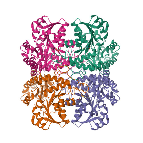
Crystal structure of cystathionine gamma synthase from Xanthomonas oryzae pv. oryzae in complex with aminoacrylate and cysteine
Ngo, H.P.T., Nguyen, T.D.Q., Kang, L.W.To be published.
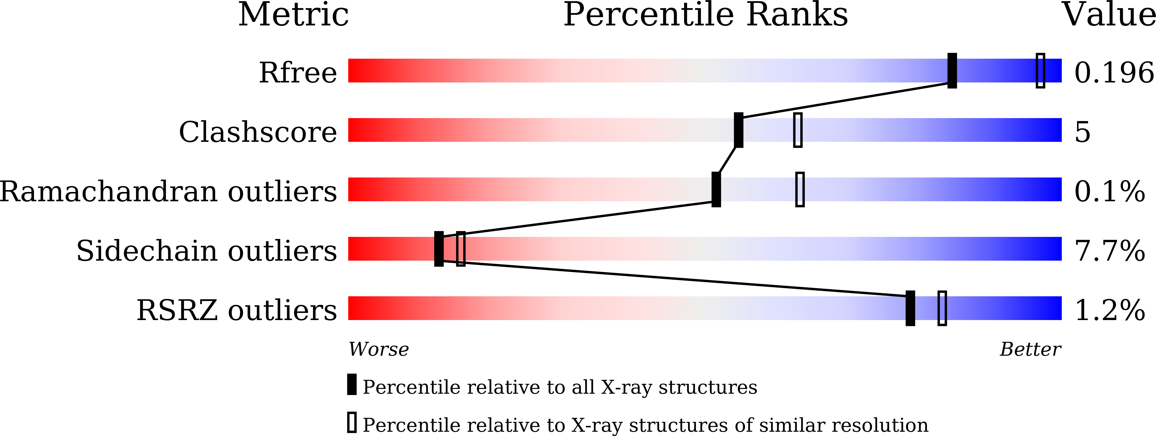
Experimental Data Snapshot
Starting Model: experimental
View more details
Entity ID: 1 | |||||
|---|---|---|---|---|---|
| Molecule | Chains | Sequence Length | Organism | Details | Image |
| Cystathionine gamma-synthase | 408 | Xanthomonas oryzae pv. oryzae KACC 10331 | Mutation(s): 1 Gene Names: metB, XOO1818 |  | |
UniProt | |||||
Find proteins for Q5H1U9 (Xanthomonas oryzae pv. oryzae (strain KACC10331 / KXO85)) Explore Q5H1U9 Go to UniProtKB: Q5H1U9 | |||||
Entity Groups | |||||
| Sequence Clusters | 30% Identity50% Identity70% Identity90% Identity95% Identity100% Identity | ||||
| UniProt Group | Q5H1U9 | ||||
Sequence AnnotationsExpand | |||||
| |||||
| Ligands 2 Unique | |||||
|---|---|---|---|---|---|
| ID | Chains | Name / Formula / InChI Key | 2D Diagram | 3D Interactions | |
| 0JO (Subject of Investigation/LOI) Query on 0JO | E [auth A], G [auth B], I [auth C], K [auth D] | 2-{[(E)-{3-hydroxy-2-methyl-5-[(phosphonooxy)methyl]pyridin-4-yl}methylidene]amino}prop-2-enoic acid C11 H13 N2 O7 P BHIGINKEEFZJGX-YIXHJXPBSA-N |  | ||
| CYS (Subject of Investigation/LOI) Query on CYS | F [auth A], H [auth B], J [auth C], L [auth D] | CYSTEINE C3 H7 N O2 S XUJNEKJLAYXESH-REOHCLBHSA-N |  | ||
| Length ( Å ) | Angle ( ˚ ) |
|---|---|
| a = 161.985 | α = 90 |
| b = 161.985 | β = 90 |
| c = 245.18 | γ = 90 |
| Software Name | Purpose |
|---|---|
| REFMAC | refinement |
| HKL-2000 | data reduction |
| PDB_EXTRACT | data extraction |
| HKL-2000 | data scaling |
| PHASES | phasing |