6LGI | pdb_00006lgi

Bombyx mori GH13 sucrose hydrolase mutant E322Q covalent intermediate complexed with fructose


Experimental Data Snapshot

  • Method: X-RAY DIFFRACTION
  • Resolution: 1.60 Å
  • R-Value Free: 
    0.207 (Depositor), 0.220 (DCC) 
  • R-Value Work: 
    0.178 (Depositor), 0.190 (DCC) 
  • R-Value Observed: 
    0.179 (Depositor) 

Starting Model: experimental
View more details

wwPDB Validation   3D Report Full Report


Ligand Structure Quality Assessment 

Created with Raphaël 2.3.0Worse 01 BetterLigand structure goodness of fit to experimental dataBest fitted BDFClick on this verticalbar to view detailsBest fitted FRUClick on this verticalbar to view detailsBest fitted BGCClick on this verticalbar to view details

This is version 1.4 of the entry. See complete history


Literature

Structure-function analysis of silkworm sucrose hydrolase uncovers the mechanism of substrate specificity in GH13 subfamily 17exo-alpha-glucosidases.

Miyazaki, T.Park, E.Y.

(2020) J Biological Chem 295: 8784-8797

  • DOI: https://doi.org/10.1074/jbc.RA120.013595
  • Primary Citation of Related Structures:  
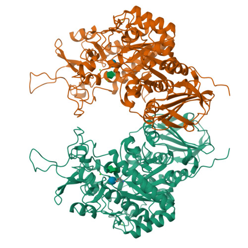
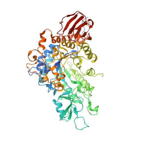
    6LGA, 6LGB, 6LGC, 6LGD, 6LGE, 6LGF, 6LGG, 6LGH, 6LGI

  • PubMed Abstract: 

    The domestic silkworm Bombyx mori expresses two sucrose-hydrolyzing enzymes, BmSUH and BmSUC1, belonging to glycoside hydrolase family 13 subfamily 17 (GH13_17) and GH32, respectively. BmSUH has little activity on maltooligosaccharides, whereas other insect GH13_17 α-glucosidases are active on sucrose and maltooligosaccharides. Little is currently known about the structural mechanisms and substrate specificity of GH13_17 enzymes. In this study, we examined the crystal structures of BmSUH without ligands; in complexes with substrates, products, and inhibitors; and complexed with its covalent intermediate at 1.60-1.85 Å resolutions. These structures revealed that the conformations of amino acid residues around subsite -1 are notably different at each step of the hydrolytic reaction. Such changes have not been previously reported among GH13 enzymes, including exo - and endo -acting hydrolases, such as α-glucosidases and α-amylases. Amino acid residues at subsite +1 are not conserved in BmSUH and other GH13_17 α-glucosidases, but subsite -1 residues are absolutely conserved. Substitutions in three subsite +1 residues, Gln 191 , Tyr 251 , and Glu 440 , decreased sucrose hydrolysis and increased maltase activity of BmSUH, indicating that these residues are key for determining its substrate specificity. These results provide detailed insights into structure-function relationships in GH13 enzymes and into the molecular evolution of insect GH13_17 α-glucosidases.


  • Organizational Affiliation

    Green Chemistry Research Division, Research Institute of Green Science and Technology, Shizuoka University, Shizuoka, Japan. Electronic address: miyazaki.takatsugu@shizuoka.ac.jp.


Macromolecules
Find similar proteins by:  (by identity cutoff)  |  3D Structure
Entity ID: 1
MoleculeChains Sequence LengthOrganismDetailsImage
Sucrose hydrolase
A, B
598Bombyx moriMutation(s): 1 
Gene Names: BmSuh
EC: 3.2.1.20
UniProt
Find proteins for A0A077JI83 (Bombyx mori)
Explore A0A077JI83 
Go to UniProtKB:  A0A077JI83
Entity Groups  
Sequence Clusters30% Identity50% Identity70% Identity90% Identity95% Identity100% Identity
UniProt GroupA0A077JI83
Sequence Annotations
Expand
  • Reference Sequence
Small Molecules
Ligands 6 Unique
IDChains Name / Formula / InChI Key2D Diagram3D Interactions
BDF (Subject of Investigation/LOI)
Query on BDF

Download Ideal Coordinates CCD File 
H [auth A],
P [auth B]
beta-D-fructopyranose
C6 H12 O6
LKDRXBCSQODPBY-ARQDHWQXSA-N
FRU (Subject of Investigation/LOI)
Query on FRU

Download Ideal Coordinates CCD File 
G [auth A],
O [auth B]
beta-D-fructofuranose
C6 H12 O6
RFSUNEUAIZKAJO-ARQDHWQXSA-N
BGC (Subject of Investigation/LOI)
Query on BGC

Download Ideal Coordinates CCD File 
F [auth A],
N [auth B]
beta-D-glucopyranose
C6 H12 O6
WQZGKKKJIJFFOK-VFUOTHLCSA-N
ACT
Query on ACT

Download Ideal Coordinates CCD File 
I [auth A],
Q [auth B]
ACETATE ION
C2 H3 O2
QTBSBXVTEAMEQO-UHFFFAOYSA-M
CA (Subject of Investigation/LOI)
Query on CA

Download Ideal Coordinates CCD File 
D [auth A],
L [auth B]
CALCIUM ION
Ca
BHPQYMZQTOCNFJ-UHFFFAOYSA-N
MG (Subject of Investigation/LOI)
Query on MG

Download Ideal Coordinates CCD File 
C [auth A],
E [auth A],
J [auth A],
K [auth B],
M [auth B]
MAGNESIUM ION
Mg
JLVVSXFLKOJNIY-UHFFFAOYSA-N
Experimental Data & Validation

Experimental Data

  • Method: X-RAY DIFFRACTION
  • Resolution: 1.60 Å
  • R-Value Free:  0.207 (Depositor), 0.220 (DCC) 
  • R-Value Work:  0.178 (Depositor), 0.190 (DCC) 
  • R-Value Observed: 0.179 (Depositor) 
Space Group: P 21 21 21
Unit Cell:
Length ( Å )Angle ( ˚ )
a = 65.405α = 90
b = 147.055β = 90
c = 153.544γ = 90
Software Package:
Software NamePurpose
REFMACrefinement
PDB_EXTRACTdata extraction
XDSdata reduction
XDSdata scaling
MOLREPphasing

Structure Validation

View Full Validation Report


Ligand Structure Quality Assessment 

Created with Raphaël 2.3.0Worse 01 BetterLigand structure goodness of fit to experimental dataBest fitted BDFClick on this verticalbar to view detailsBest fitted FRUClick on this verticalbar to view detailsBest fitted BGCClick on this verticalbar to view details

Entry History & Funding Information

Deposition Data

  • Released Date: 2020-05-20 
  • Deposition Author(s): Miyazaki, T.

Funding OrganizationLocationGrant Number
Japan Society for the Promotion of ScienceJapan19K15748

Revision History  (Full details and data files)

  • Version 1.0: 2020-05-20
    Type: Initial release
  • Version 1.1: 2020-07-08
    Changes: Database references
  • Version 1.2: 2020-07-29
    Type: Remediation
    Reason: Carbohydrate remediation
    Changes: Data collection, Derived calculations, Structure summary
  • Version 1.3: 2023-11-22
    Changes: Data collection, Database references, Refinement description, Structure summary
  • Version 1.4: 2024-11-06
    Changes: Structure summary