6N4I

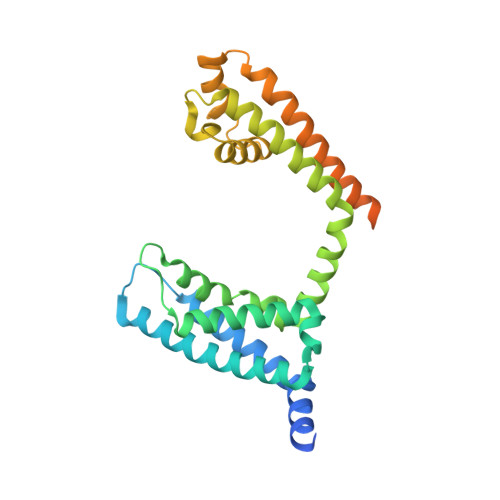
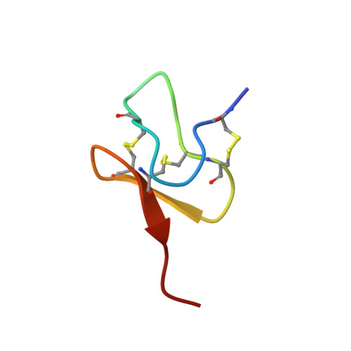
Structural basis of Nav1.7 inhibition by a gating-modifier spider toxin


Experimental Data Snapshot

  • Method: X-RAY DIFFRACTION
  • Resolution: 3.54 Å
  • R-Value Free: 0.294 
  • R-Value Work: 0.275 
  • R-Value Observed: 0.276 

Starting Model: experimental
View more details

wwPDB Validation   3D Report Full Report


Ligand Structure Quality Assessment 


This is version 1.4 of the entry. See complete history


Literature

Structural Basis of Nav1.7 Inhibition by a Gating-Modifier Spider Toxin.

Xu, H.Li, T.Rohou, A.Arthur, C.P.Tzakoniati, F.Wong, E.Estevez, A.Kugel, C.Franke, Y.Chen, J.Ciferri, C.Hackos, D.H.Koth, C.M.Payandeh, J.

(2019) Cell 176: 702-715.e14

  • DOI: https://doi.org/10.1016/j.cell.2018.12.018
  • Primary Citation of Related Structures:  
    6N4I, 6N4Q, 6N4R

  • PubMed Abstract: 

    Voltage-gated sodium (Nav) channels are targets of disease mutations, toxins, and therapeutic drugs. Despite recent advances, the structural basis of voltage sensing, electromechanical coupling, and toxin modulation remains ill-defined. Protoxin-II (ProTx2) from the Peruvian green velvet tarantula is an inhibitor cystine-knot peptide and selective antagonist of the human Nav1.7 channel. Here, we visualize ProTx2 in complex with voltage-sensor domain II (VSD2) from Nav1.7 using X-ray crystallography and cryoelectron microscopy. Membrane partitioning orients ProTx2 for unfettered access to VSD2, where ProTx2 interrogates distinct features of the Nav1.7 receptor site. ProTx2 positions two basic residues into the extracellular vestibule to antagonize S4 gating-charge movement through an electrostatic mechanism. ProTx2 has trapped activated and deactivated states of VSD2, revealing a remarkable ∼10 Å translation of the S4 helix, providing a structural framework for activation gating in voltage-gated ion channels. Finally, our results deliver key templates to design selective Nav channel antagonists.


  • Organizational Affiliation

    Department of Structural Biology, Genentech, South San Francisco, CA 94080, USA.


Macromolecules
Find similar proteins by:  (by identity cutoff)  |  3D Structure
Entity ID: 1
MoleculeChains Sequence LengthOrganismDetailsImage
Nav1.7 VSD2-NavAb channel chimera protein
A, B, C, D
288Homo sapiensMutation(s): 0 
Entity Groups  
Sequence Clusters30% Identity50% Identity70% Identity90% Identity95% Identity100% Identity
Sequence Annotations
Expand
  • Reference Sequence
Find similar proteins by:  (by identity cutoff)  |  3D Structure
Entity ID: 2
MoleculeChains Sequence LengthOrganismDetailsImage
Beta/omega-theraphotoxin-Tp2a
E, F, G, H
30Thrixopelma pruriensMutation(s): 0 
UniProt
Find proteins for P83476 (Thrixopelma pruriens)
Explore P83476 
Go to UniProtKB:  P83476
Entity Groups  
Sequence Clusters30% Identity50% Identity70% Identity90% Identity95% Identity100% Identity
UniProt GroupP83476
Sequence Annotations
Expand
  • Reference Sequence
Small Molecules
Ligands 1 Unique
IDChains Name / Formula / InChI Key2D Diagram3D Interactions
6OU
Query on 6OU

Download Ideal Coordinates CCD File 
I [auth A]
J [auth A]
K [auth A]
L [auth B]
M [auth B]
I [auth A],
J [auth A],
K [auth A],
L [auth B],
M [auth B],
N [auth B],
O [auth C],
P [auth C],
Q [auth D],
R [auth D],
S [auth D],
T [auth D],
U [auth D],
V [auth E],
W [auth F],
X [auth G]
[(2~{R})-1-[2-azanylethoxy(oxidanyl)phosphoryl]oxy-3-hexadecanoyloxy-propan-2-yl] (~{Z})-octadec-9-enoate
C39 H76 N O8 P
FHQVHHIBKUMWTI-OTMQOFQLSA-N
Experimental Data & Validation

Experimental Data

  • Method: X-RAY DIFFRACTION
  • Resolution: 3.54 Å
  • R-Value Free: 0.294 
  • R-Value Work: 0.275 
  • R-Value Observed: 0.276 
  • Space Group: C 1 2 1
Unit Cell:
Length ( Å )Angle ( ˚ )
a = 221.22α = 90
b = 123.53β = 124
c = 123.99γ = 90
Software Package:
Software NamePurpose
PHENIXrefinement
XDSdata reduction
XSCALEdata scaling
PHENIXphasing

Structure Validation

View Full Validation Report


Ligand Structure Quality Assessment 


Entry History 

Deposition Data

Revision History  (Full details and data files)

  • Version 1.0: 2019-01-23
    Type: Initial release
  • Version 1.1: 2019-02-06
    Changes: Data collection, Database references, Structure summary
  • Version 1.2: 2019-02-20
    Changes: Data collection, Database references
  • Version 1.3: 2023-10-11
    Changes: Data collection, Database references, Refinement description
  • Version 1.4: 2024-11-20
    Changes: Structure summary