6N4L | pdb_00006n4l

Dithionite-reduced ADP-bound form of the nitrogenase Fe-protein from A. vinelandii


Experimental Data Snapshot

  • Method: X-RAY DIFFRACTION
  • Resolution: 1.13 Å
  • R-Value Free: 
    0.157 (Depositor), 0.150 (DCC) 
  • R-Value Work: 
    0.146 (Depositor), 0.150 (DCC) 
  • R-Value Observed: 
    0.146 (Depositor) 

Starting Model: experimental
View more details

wwPDB Validation   3D Report Full Report


Ligand Structure Quality Assessment 

Created with Raphaël 2.3.0Worse 01 BetterLigand structure goodness of fit to experimental dataBest fitted ADPClick on this verticalbar to view details

This is version 1.4 of the entry. See complete history


Literature

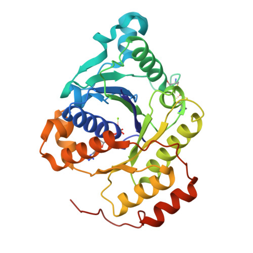
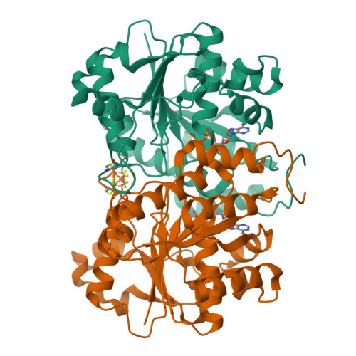
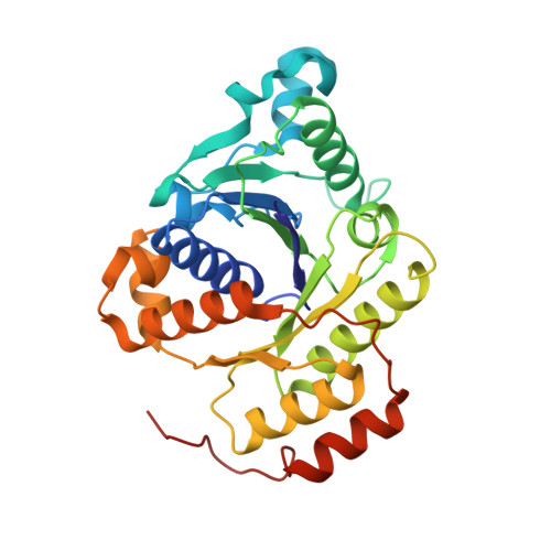
Site-Specific Oxidation State Assignments of the Iron Atoms in the [4Fe:4S]2+/1+/0States of the Nitrogenase Fe-Protein.

Wenke, B.B.Spatzal, T.Rees, D.C.

(2019) Angew Chem Int Ed Engl 58: 3894-3897

  • DOI: https://doi.org/10.1002/anie.201813966
  • Primary Citation of Related Structures:  
    6N4J, 6N4K, 6N4L, 6N4M

  • PubMed Abstract: 

    The nitrogenase iron protein (Fe-protein) contains an unusual [4Fe:4S] iron-sulphur cluster that is stable in three oxidation states: 2+, 1+, and 0. Here, we use spatially resolved anomalous dispersion (SpReAD) refinement to determine oxidation assignments for the individual irons for each state. Additionally, we report the 1.13-Å resolution structure for the ADP bound Fe-protein, the highest resolution Fe-protein structure presently determined. In the dithionite-reduced [4Fe:4S] 1+ state, our analysis identifies a solvent exposed, delocalized Fe 2.5+ pair and a buried Fe 2+ pair. We propose that ATP binding by the Fe-protein promotes an internal redox rearrangement such that the solvent-exposed Fe pair becomes reduced, thereby facilitating electron transfer to the nitrogenase molybdenum iron-protein. In the [4Fe:4S] 0 and [4Fe:4S] 2+ states, the SpReAD analysis supports oxidation states assignments for all irons in these clusters of Fe 2+ and valence delocalized Fe 2.5+ , respectively.


  • Organizational Affiliation

    Division of Chemistry and Chemical Engineering, California Institute of Technology, Pasadena, CA, 91125, USA.


Macromolecules
Find similar proteins by:  (by identity cutoff)  |  3D Structure
Entity ID: 1
MoleculeChains Sequence LengthOrganismDetailsImage
Nitrogenase iron protein 1289Azotobacter vinelandiiMutation(s): 0 
EC: 1.18.6.1
UniProt
Find proteins for P00459 (Azotobacter vinelandii)
Explore P00459 
Go to UniProtKB:  P00459
Entity Groups  
Sequence Clusters30% Identity50% Identity70% Identity90% Identity95% Identity100% Identity
UniProt GroupP00459
Sequence Annotations
Expand
  • Reference Sequence
Experimental Data & Validation

Experimental Data

  • Method: X-RAY DIFFRACTION
  • Resolution: 1.13 Å
  • R-Value Free:  0.157 (Depositor), 0.150 (DCC) 
  • R-Value Work:  0.146 (Depositor), 0.150 (DCC) 
  • R-Value Observed: 0.146 (Depositor) 
Space Group: P 2 21 21
Unit Cell:
Length ( Å )Angle ( ˚ )
a = 45.587α = 90
b = 74.63β = 90
c = 75.398γ = 90
Software Package:
Software NamePurpose
REFMACrefinement
SCALAdata scaling
PDB_EXTRACTdata extraction
XDSdata reduction
PHASERphasing

Structure Validation

View Full Validation Report


Ligand Structure Quality Assessment 

Created with Raphaël 2.3.0Worse 01 BetterLigand structure goodness of fit to experimental dataBest fitted ADPClick on this verticalbar to view details

Entry History & Funding Information

Deposition Data


Funding OrganizationLocationGrant Number
National Institutes of Health/National Institute of General Medical Sciences (NIH/NIGMS)United StatesGM045162

Revision History  (Full details and data files)

  • Version 1.0: 2019-02-13
    Type: Initial release
  • Version 1.1: 2019-03-27
    Changes: Data collection, Database references
  • Version 1.2: 2020-01-01
    Changes: Author supporting evidence
  • Version 1.3: 2023-10-11
    Changes: Data collection, Database references, Derived calculations, Refinement description
  • Version 1.4: 2024-11-06
    Changes: Structure summary