6PW1

Cytochrome c Oxidase delta 16


Experimental Data Snapshot

  • Method: X-RAY DIFFRACTION
  • Resolution: 2.10 Å
  • R-Value Free: 0.204 
  • R-Value Work: 0.180 
  • R-Value Observed: 0.180 

Starting Model: experimental
View more details

wwPDB Validation   3D Report Full Report


Ligand Structure Quality Assessment 


This is version 2.1 of the entry. See complete history


Literature

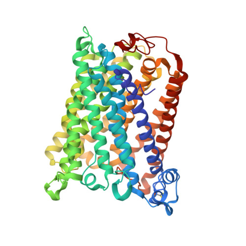
Structural changes at the surface of cytochrome c oxidase alter the proton-pumping stoichiometry.

Berg, J.Liu, J.Svahn, E.Ferguson-Miller, S.Brzezinski, P.

(2019) Biochim Biophys Acta Bioenerg 1861: 148116-148116

  • DOI: https://doi.org/10.1016/j.bbabio.2019.148116
  • Primary Citation of Related Structures:  
    6PW0, 6PW1

  • PubMed Abstract: 

    Data from earlier studies showed that minor structural changes at the surface of cytochrome c oxidase, in one of the proton-input pathways (the D pathway), result in dramatically decreased activity and a lower proton-pumping stoichiometry. To further investigate how changes around the D pathway orifice influence functionality of the enzyme, here we modified the nearby C-terminal loop of subunit I of the Rhodobacter sphaeroides cytochrome c oxidase. Removal of 16 residues from this flexible surface loop resulted in a decrease in the proton-pumping stoichiometry to <50% of that of the wild-type enzyme. Replacement of the protonatable residue Glu552, part of the same loop, by an Ala, resulted in a similar decrease in the proton-pumping stoichiometry without loss of the O 2 -reduction activity or changes in the proton-uptake kinetics. The data show that minor structural changes at the orifice of the D pathway, at a distance of ~40 Å from the proton gate of cytochrome c oxidase, may alter the proton-pumping stoichiometry of the enzyme.


  • Organizational Affiliation

    Department of Biochemistry and Biophysics, The Arrhenius Laboratories for Natural Sciences, Stockholm University, SE-106 91 Stockholm, Sweden.


Macromolecules
Find similar proteins by:  (by identity cutoff)  |  3D Structure
Entity ID: 1
MoleculeChains Sequence LengthOrganismDetailsImage
Cytochrome c oxidase subunit 1
A, C
534Cereibacter sphaeroides 2.4.1Mutation(s): 0 
Gene Names: coxIRSP_1877
EC: 1.9.3.1
Membrane Entity: Yes 
UniProt
Find proteins for Q3J5A7 (Cereibacter sphaeroides (strain ATCC 17023 / DSM 158 / JCM 6121 / CCUG 31486 / LMG 2827 / NBRC 12203 / NCIMB 8253 / ATH 2.4.1.))
Explore Q3J5A7 
Go to UniProtKB:  Q3J5A7
Entity Groups  
Sequence Clusters30% Identity50% Identity70% Identity90% Identity95% Identity100% Identity
UniProt GroupQ3J5A7
Sequence Annotations
Expand
  • Reference Sequence
Find similar proteins by:  (by identity cutoff)  |  3D Structure
Entity ID: 2
MoleculeChains Sequence LengthOrganismDetailsImage
Cytochrome c oxidase subunit 2
B, D
257Cereibacter sphaeroides 2.4.1Mutation(s): 0 
Gene Names: coxIIRSP_1826
EC: 1.9.3.1
Membrane Entity: Yes 
UniProt
Find proteins for Q3J5G0 (Cereibacter sphaeroides (strain ATCC 17023 / DSM 158 / JCM 6121 / CCUG 31486 / LMG 2827 / NBRC 12203 / NCIMB 8253 / ATH 2.4.1.))
Explore Q3J5G0 
Go to UniProtKB:  Q3J5G0
Entity Groups  
Sequence Clusters30% Identity50% Identity70% Identity90% Identity95% Identity100% Identity
UniProt GroupQ3J5G0
Sequence Annotations
Expand
  • Reference Sequence
Oligosaccharides

Help

Entity ID: 3
MoleculeChains Length2D Diagram Glycosylation3D Interactions
alpha-D-glucopyranose-(1-4)-alpha-D-glucopyranose
E, F
2N/A
Glycosylation Resources
GlyTouCan:  G07411ON
GlyCosmos:  G07411ON
Small Molecules
Ligands 9 Unique
IDChains Name / Formula / InChI Key2D Diagram3D Interactions
HEA (Subject of Investigation/LOI)
Query on HEA

Download Ideal Coordinates CCD File 
LA [auth C],
MA [auth C],
U [auth A],
V [auth A]
HEME-A
C49 H56 Fe N4 O6
ZGGYGTCPXNDTRV-PRYGPKJJSA-L
DMU
Query on DMU

Download Ideal Coordinates CCD File 
BA [auth B]
KA [auth C]
Q [auth A]
R [auth A]
S [auth A]
BA [auth B],
KA [auth C],
Q [auth A],
R [auth A],
S [auth A],
T [auth A]
DECYL-BETA-D-MALTOPYRANOSIDE
C22 H42 O11
WOQQAWHSKSSAGF-WXFJLFHKSA-N
TRD
Query on TRD

Download Ideal Coordinates CCD File 
AA [auth B]
G [auth A]
H [auth A]
I [auth A]
IA [auth C]
AA [auth B],
G [auth A],
H [auth A],
I [auth A],
IA [auth C],
J [auth A],
JA [auth C],
K [auth A],
L [auth A],
M [auth A],
N [auth A],
O [auth A],
P [auth A],
QA [auth D]
TRIDECANE
C13 H28
IIYFAKIEWZDVMP-UHFFFAOYSA-N
HTH
Query on HTH

Download Ideal Coordinates CCD File 
HA [auth B],
Z [auth A]
(2S,3R)-heptane-1,2,3-triol
C7 H16 O3
HXYCHJFUBNTKQR-RQJHMYQMSA-N
TRS
Query on TRS

Download Ideal Coordinates CCD File 
CA [auth B]2-AMINO-2-HYDROXYMETHYL-PROPANE-1,3-DIOL
C4 H12 N O3
LENZDBCJOHFCAS-UHFFFAOYSA-O
CD
Query on CD

Download Ideal Coordinates CCD File 
FA [auth B],
GA [auth B],
TA [auth D],
UA [auth D]
CADMIUM ION
Cd
WLZRMCYVCSSEQC-UHFFFAOYSA-N
CU
Query on CU

Download Ideal Coordinates CCD File 
DA [auth B]
EA [auth B]
NA [auth C]
RA [auth D]
SA [auth D]
DA [auth B],
EA [auth B],
NA [auth C],
RA [auth D],
SA [auth D],
W [auth A]
COPPER (II) ION
Cu
JPVYNHNXODAKFH-UHFFFAOYSA-N
CA
Query on CA

Download Ideal Coordinates CCD File 
PA [auth C],
Y [auth A]
CALCIUM ION
Ca
BHPQYMZQTOCNFJ-UHFFFAOYSA-N
MG
Query on MG

Download Ideal Coordinates CCD File 
OA [auth C],
X [auth A]
MAGNESIUM ION
Mg
JLVVSXFLKOJNIY-UHFFFAOYSA-N
Biologically Interesting Molecules (External Reference) 1 Unique
Experimental Data & Validation

Experimental Data

  • Method: X-RAY DIFFRACTION
  • Resolution: 2.10 Å
  • R-Value Free: 0.204 
  • R-Value Work: 0.180 
  • R-Value Observed: 0.180 
  • Space Group: P 21 21 21
Unit Cell:
Length ( Å )Angle ( ˚ )
a = 123.315α = 90
b = 130β = 90
c = 177.539γ = 90
Software Package:
Software NamePurpose
PHENIXrefinement
HKL-2000data scaling
PHASERphasing
HKL-2000data reduction

Structure Validation

View Full Validation Report


Ligand Structure Quality Assessment 


Entry History & Funding Information

Deposition Data


Funding OrganizationLocationGrant Number
National Institutes of Health/National Institute of General Medical Sciences (NIH/NIGMS)United States--

Revision History  (Full details and data files)

  • Version 1.0: 2019-11-27
    Type: Initial release
  • Version 1.1: 2019-12-11
    Changes: Database references
  • Version 1.2: 2020-01-01
    Changes: Author supporting evidence
  • Version 2.0: 2020-07-29
    Type: Remediation
    Reason: Carbohydrate remediation
    Changes: Advisory, Atomic model, Data collection, Derived calculations, Non-polymer description, Structure summary
  • Version 2.1: 2023-10-11
    Changes: Advisory, Data collection, Database references, Refinement description, Structure summary