6Q39 | pdb_00006q39

Complex of Arginase 2 with Example 49


Experimental Data Snapshot

  • Method: X-RAY DIFFRACTION
  • Resolution: 2.21 Å
  • R-Value Free: 
    0.210 (Depositor), 0.170 (DCC) 
  • R-Value Work: 
    0.162 (Depositor), 0.160 (DCC) 
  • R-Value Observed: 
    0.165 (Depositor) 

Starting Model: experimental
View more details

wwPDB Validation   3D Report Full Report


Ligand Structure Quality Assessment 

Created with Raphaël 2.3.0Worse 01 BetterLigand structure goodness of fit to experimental dataBest fitted HDQClick on this verticalbar to view details

This is version 2.1 of the entry. See complete history


Literature

Discovery ofN-Substituted 3-Amino-4-(3-boronopropyl)pyrrolidine-3-carboxylic Acids as Highly Potent Third-Generation Inhibitors of Human Arginase I and II.

Van Zandt, M.C.Jagdmann, G.E.Whitehouse, D.L.Ji, M.Savoy, J.Potapova, O.Cousido-Siah, A.Mitschler, A.Howard, E.I.Pyle, A.M.Podjarny, A.D.

(2019) J Med Chem 62: 8164-8177

  • DOI: https://doi.org/10.1021/acs.jmedchem.9b00931
  • Primary Citation of Related Structures:  
    6Q37, 6Q39

  • PubMed Abstract: 

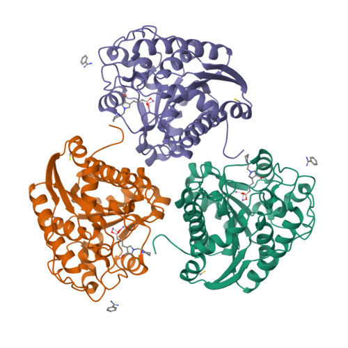
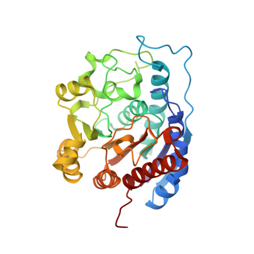
    Recent efforts to identify new highly potent arginase inhibitors have resulted in the discovery of a novel family of (3 R ,4 S )-3-amino-4-(3-boronopropyl)pyrrolidine-3-carboxylic acid analogues with up to a 1000-fold increase in potency relative to the current standards, 2-amino-6-boronohexanoic acid (ABH) and N -hydroxy-nor-l-arginine (nor-NOHA). The lead candidate, with an N -2-amino-3-phenylpropyl substituent (NED-3238), example 43 , inhibits arginase I and II with IC 50 values of 1.3 and 8.1 nM, respectively. Herein, we report the design, synthesis, and structure-activity relationships for this novel series of inhibitors, along with X-ray crystallographic data for selected examples bound to human arginase II.


  • Organizational Affiliation

    New England Discovery Partners , 23 Business Park Drive , Branford , Connecticut 06405 , United States.


Macromolecules
Find similar proteins by:  (by identity cutoff)  |  3D Structure
Entity ID: 1
MoleculeChains Sequence LengthOrganismDetailsImage
Arginase-2, mitochondrial
A, B, C
306Homo sapiensMutation(s): 0 
Gene Names: ARG2
EC: 3.5.3.1
UniProt & NIH Common Fund Data Resources
Find proteins for P78540 (Homo sapiens)
Explore P78540 
Go to UniProtKB:  P78540
PHAROS:  P78540
GTEx:  ENSG00000081181 
Entity Groups  
Sequence Clusters30% Identity50% Identity70% Identity90% Identity95% Identity100% Identity
UniProt GroupP78540
Sequence Annotations
Expand
  • Reference Sequence
Small Molecules
Ligands 4 Unique
IDChains Name / Formula / InChI Key2D Diagram3D Interactions
HDQ (Subject of Investigation/LOI)
Query on HDQ

Download Ideal Coordinates CCD File 
H [auth A],
M [auth B],
R [auth C]
3-[(3~{S},4~{R})-4-azanyl-4-carboxy-1-[[(2~{S})-piperidin-2-yl]methyl]pyrrolidin-3-yl]propyl-tris(oxidanyl)boranuide
C14 H29 B N3 O5
UAQSNVVPOJZDTG-OBJOEFQTSA-N
BEN
Query on BEN

Download Ideal Coordinates CCD File 
F [auth A],
K [auth B],
P [auth C]
BENZAMIDINE
C7 H8 N2
PXXJHWLDUBFPOL-UHFFFAOYSA-N
BME
Query on BME

Download Ideal Coordinates CCD File 
G [auth A],
L [auth B],
Q [auth C]
BETA-MERCAPTOETHANOL
C2 H6 O S
DGVVWUTYPXICAM-UHFFFAOYSA-N
MN
Query on MN

Download Ideal Coordinates CCD File 
D [auth A]
E [auth A]
I [auth B]
J [auth B]
N [auth C]
MANGANESE (II) ION
Mn
WAEMQWOKJMHJLA-UHFFFAOYSA-N
Experimental Data & Validation

Experimental Data

  • Method: X-RAY DIFFRACTION
  • Resolution: 2.21 Å
  • R-Value Free:  0.210 (Depositor), 0.170 (DCC) 
  • R-Value Work:  0.162 (Depositor), 0.160 (DCC) 
  • R-Value Observed: 0.165 (Depositor) 
Space Group: P 42 21 2
Unit Cell:
Length ( Å )Angle ( ˚ )
a = 128.303α = 90
b = 128.303β = 90
c = 159.155γ = 90
Software Package:
Software NamePurpose
PHENIXrefinement
SCALEPACKdata scaling
PDB_EXTRACTdata extraction
DENZOdata reduction
AMoREphasing

Structure Validation

View Full Validation Report


Ligand Structure Quality Assessment 

Created with Raphaël 2.3.0Worse 01 BetterLigand structure goodness of fit to experimental dataBest fitted HDQClick on this verticalbar to view details

Entry History 

Deposition Data

Revision History  (Full details and data files)

  • Version 1.0: 2019-08-28
    Type: Initial release
  • Version 1.1: 2019-09-25
    Changes: Data collection, Database references
  • Version 1.2: 2019-10-16
    Changes: Data collection
  • Version 2.0: 2021-08-04
    Changes: Atomic model, Data collection, Derived calculations, Non-polymer description, Structure summary
  • Version 2.1: 2024-01-24
    Changes: Data collection, Database references, Refinement description