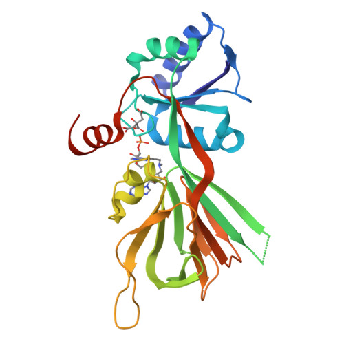
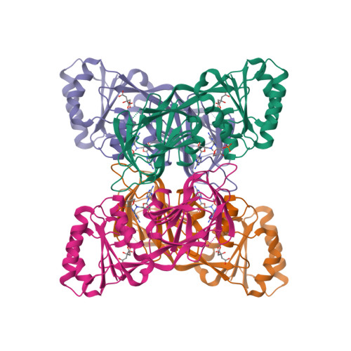
Crystal structure of NAD kinase 1 from Listeria monocytogenes in complex with an inhibitor
Gelin, M., Labesse, G.To be published.
Experimental Data Snapshot
Entity ID: 1 | |||||
|---|---|---|---|---|---|
| Molecule | Chains | Sequence Length | Organism | Details | Image |
| NAD kinase 1 | 272 | Listeria monocytogenes EGD-e | Mutation(s): 0 Gene Names: nadK1, lmo0968 EC: 2.7.1.23 | 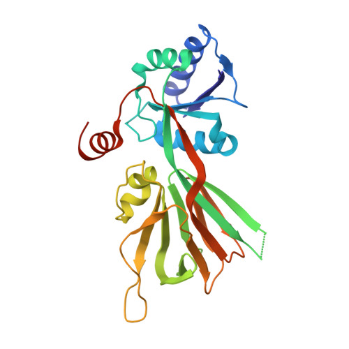 | |
UniProt | |||||
Find proteins for Q8Y8D7 (Listeria monocytogenes serovar 1/2a (strain ATCC BAA-679 / EGD-e)) Explore Q8Y8D7 Go to UniProtKB: Q8Y8D7 | |||||
Entity Groups | |||||
| Sequence Clusters | 30% Identity50% Identity70% Identity90% Identity95% Identity100% Identity | ||||
| UniProt Group | Q8Y8D7 | ||||
Sequence AnnotationsExpand | |||||
| |||||
| Ligands 2 Unique | |||||
|---|---|---|---|---|---|
| ID | Chains | Name / Formula / InChI Key | 2D Diagram | 3D Interactions | |
| K28 Query on K28 | C [auth A] | [(2~{R},3~{R},4~{R},5~{R})-2-[8-[3-[[(2~{R},3~{S},4~{R},5~{R})-5-(6-aminopurin-9-yl)-3,4-bis(oxidanyl)oxolan-2-yl]methyl-methyl-amino]prop-1-ynyl]-6-azanyl-purin-9-yl]-5-(hydroxymethyl)-4-oxidanyl-oxolan-3-yl] dihydrogen phosphate C24 H30 N11 O10 P LZWZGJMZXMPLGZ-KRSQEUQLSA-N |  | ||
| CIT Query on CIT | B [auth A] | CITRIC ACID C6 H8 O7 KRKNYBCHXYNGOX-UHFFFAOYSA-N |  | ||
| Length ( Å ) | Angle ( ˚ ) |
|---|---|
| a = 63.15 | α = 90 |
| b = 75.17 | β = 90 |
| c = 118.59 | γ = 90 |
| Software Name | Purpose |
|---|---|
| PHENIX | refinement |
| XDS | data reduction |
| Aimless | data scaling |
| PDB_EXTRACT | data extraction |
| PHENIX | phasing |