6RNK | pdb_00006rnk

Crystal structure of a humanized (K18E, K269N) rat succinate receptor SUCNR1 (GPR91) in complex with a nanobody and antagonist NF-56-EJ40.


Experimental Data Snapshot

  • Method: X-RAY DIFFRACTION
  • Resolution: 1.94 Å
  • R-Value Free: 
    0.199 (Depositor), 0.190 (DCC) 
  • R-Value Work: 
    0.177 (Depositor), 0.180 (DCC) 
  • R-Value Observed: 
    0.178 (Depositor) 

Starting Model: experimental
View more details

wwPDB Validation   3D Report Full Report


Ligand Structure Quality Assessment 

Created with Raphaël 2.3.0Worse 01 BetterLigand structure goodness of fit to experimental dataBest fitted KAZClick on this verticalbar to view detailsBest fitted OLCClick on this verticalbar to view details

This is version 1.4 of the entry. See complete history


Literature

Structural basis of species-selective antagonist binding to the succinate receptor.

Haffke, M.Fehlmann, D.Rummel, G.Boivineau, J.Duckely, M.Gommermann, N.Cotesta, S.Sirockin, F.Freuler, F.Littlewood-Evans, A.Kaupmann, K.Jaakola, V.P.

(2019) Nature 574: 581-585

  • DOI: https://doi.org/10.1038/s41586-019-1663-8
  • Primary Citation of Related Structures:  
    6IBB, 6RNK

  • PubMed Abstract: 

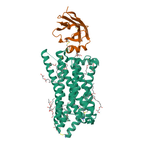
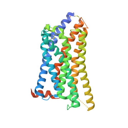
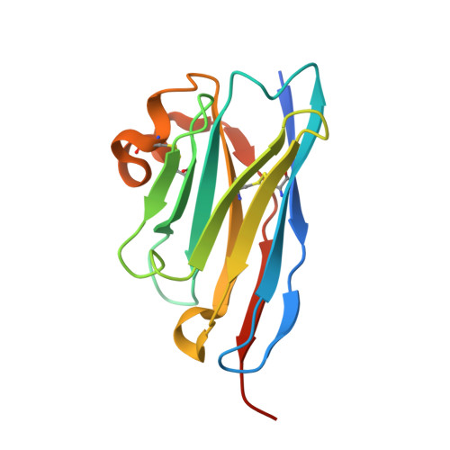
    The tricarboxylic acid cycle intermediate succinate is involved in metabolic processes and plays a crucial role in the homeostasis of mitochondrial reactive oxygen species 1 . The receptor responsible for succinate signalling, SUCNR1 (also known as GPR91), is a member of the G-protein-coupled-receptor family 2 and links succinate signalling to renin-induced hypertension, retinal angiogenesis and inflammation 3-5 . Because SUCNR1 senses succinate as an immunological danger signal 6 -which has relevance for diseases including ulcerative colitis, liver fibrosis 7 , diabetes and rheumatoid arthritis 3,8 -it is of interest as a therapeutic target. Here we report the high-resolution crystal structure of rat SUCNR1 in complex with an intracellular binding nanobody in the inactive conformation. Structure-based mutagenesis and radioligand-binding studies, in conjunction with molecular modelling, identified key residues for species-selective antagonist binding and enabled the determination of the high-resolution crystal structure of a humanized rat SUCNR1 in complex with a high-affinity, human-selective antagonist denoted NF-56-EJ40. We anticipate that these structural insights into the architecture of the succinate receptor and its antagonist selectivity will enable structure-based drug discovery and will further help to elucidate the function of SUCNR1 in vitro and in vivo.


  • Organizational Affiliation

    Chemical Biology & Therapeutics, Novartis Institutes for BioMedical Research, Novartis Pharma AG, Basel, Switzerland. matthias.haffke@novartis.com.


Macromolecules
Find similar proteins by:  (by identity cutoff)  |  3D Structure
Entity ID: 1
MoleculeChains Sequence LengthOrganismDetailsImage
Succinate receptor 1342Rattus norvegicusMutation(s): 2 
Gene Names: Sucnr1Gpr91
Membrane Entity: Yes 
UniProt
Find proteins for Q6IYF9 (Rattus norvegicus)
Explore Q6IYF9 
Go to UniProtKB:  Q6IYF9
Entity Groups  
Sequence Clusters30% Identity50% Identity70% Identity90% Identity95% Identity100% Identity
UniProt GroupQ6IYF9
Sequence Annotations
Expand
  • Reference Sequence
Find similar proteins by:  (by identity cutoff)  |  3D Structure
Entity ID: 2
MoleculeChains Sequence LengthOrganismDetailsImage
Nanobody6142Vicugna pacosMutation(s): 0 
Entity Groups  
Sequence Clusters30% Identity50% Identity70% Identity90% Identity95% Identity100% Identity
Sequence Annotations
Expand
  • Reference Sequence
Small Molecules
Ligands 4 Unique
IDChains Name / Formula / InChI Key2D Diagram3D Interactions
KAZ
Query on KAZ

Download Ideal Coordinates CCD File 
C [auth A]2-[2-[[3-[4-[(4-methylpiperazin-1-yl)methyl]phenyl]phenyl]carbonylamino]phenyl]ethanoic acid
C27 H29 N3 O3
UTWXDNZWMQAUKL-UHFFFAOYSA-N
OLC
Query on OLC

Download Ideal Coordinates CCD File 
D [auth A]
E [auth A]
F [auth A]
G [auth A]
H [auth A]
(2R)-2,3-dihydroxypropyl (9Z)-octadec-9-enoate
C21 H40 O4
RZRNAYUHWVFMIP-GDCKJWNLSA-N
SO4
Query on SO4

Download Ideal Coordinates CCD File 
R [auth A]SULFATE ION
O4 S
QAOWNCQODCNURD-UHFFFAOYSA-L
GOL
Query on GOL

Download Ideal Coordinates CCD File 
Q [auth A]GLYCEROL
C3 H8 O3
PEDCQBHIVMGVHV-UHFFFAOYSA-N
Experimental Data & Validation

Experimental Data

  • Method: X-RAY DIFFRACTION
  • Resolution: 1.94 Å
  • R-Value Free:  0.199 (Depositor), 0.190 (DCC) 
  • R-Value Work:  0.177 (Depositor), 0.180 (DCC) 
  • R-Value Observed: 0.178 (Depositor) 
Space Group: C 1 2 1
Unit Cell:
Length ( Å )Angle ( ˚ )
a = 76.83α = 90
b = 150.93β = 112.42
c = 68.23γ = 90
Software Package:
Software NamePurpose
BUSTERrefinement
XDSdata reduction
autoPROCdata scaling
PHASERphasing

Structure Validation

View Full Validation Report


Ligand Structure Quality Assessment 

Created with Raphaël 2.3.0Worse 01 BetterLigand structure goodness of fit to experimental dataBest fitted KAZClick on this verticalbar to view detailsBest fitted OLCClick on this verticalbar to view details

Entry History 

Deposition Data

Revision History  (Full details and data files)

  • Version 1.0: 2019-08-14
    Type: Initial release
  • Version 1.1: 2019-10-16
    Changes: Data collection, Database references
  • Version 1.2: 2019-11-06
    Changes: Data collection, Database references
  • Version 1.3: 2024-01-24
    Changes: Data collection, Database references, Refinement description
  • Version 1.4: 2024-11-20
    Changes: Structure summary