6RQZ

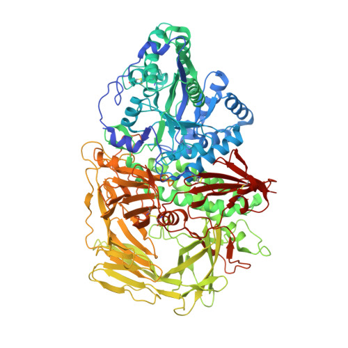
GOLGI ALPHA-MANNOSIDASE II complex with Manno-epi-cyclophellitol aziridine


Experimental Data Snapshot

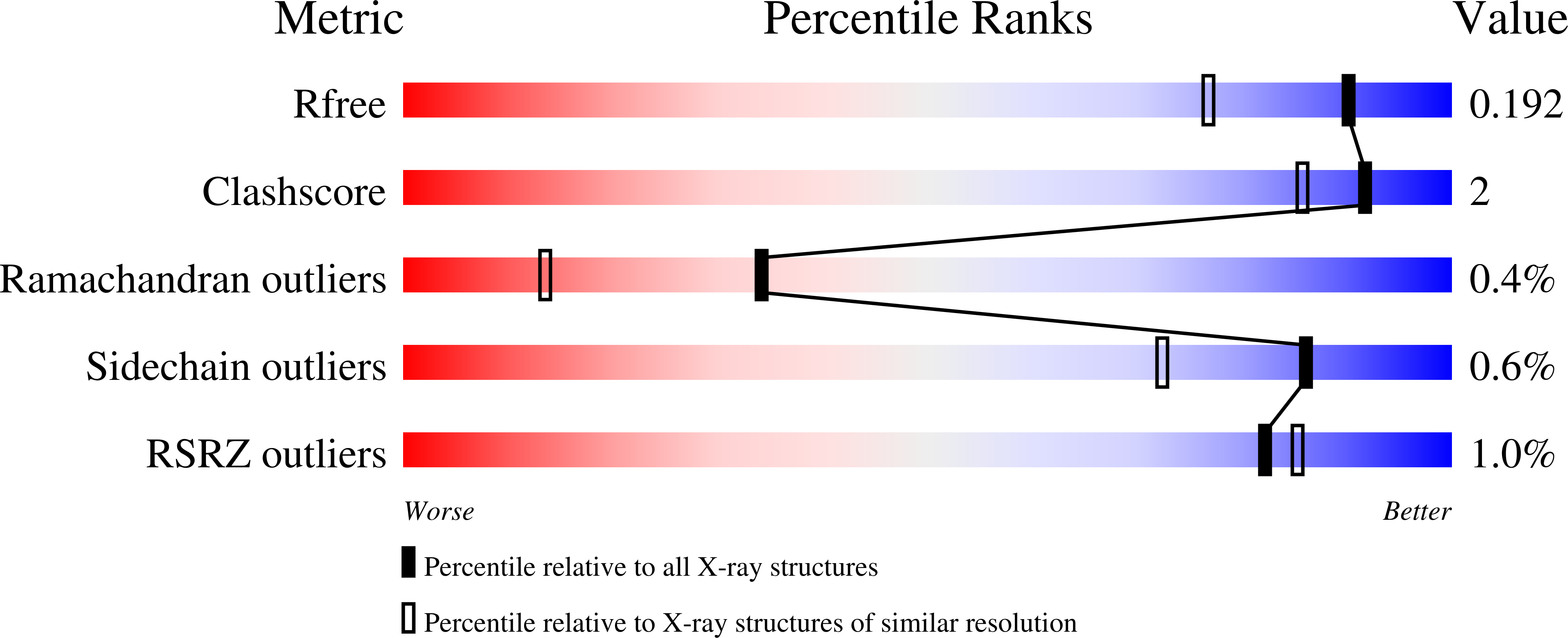
  • Method: X-RAY DIFFRACTION
  • Resolution: 1.53 Å
  • R-Value Free: 0.183 
  • R-Value Work: 0.156 
  • R-Value Observed: 0.158 

Starting Model: experimental
View more details

wwPDB Validation   3D Report Full Report


Ligand Structure Quality Assessment 


This is version 1.3 of the entry. See complete history


Literature

Manno- epi -cyclophellitols Enable Activity-Based Protein Profiling of Human alpha-Mannosidases and Discovery of New Golgi Mannosidase II Inhibitors.

Armstrong, Z.Kuo, C.L.Lahav, D.Liu, B.Johnson, R.Beenakker, T.J.M.de Boer, C.Wong, C.S.van Rijssel, E.R.Debets, M.F.Florea, B.I.Hissink, C.Boot, R.G.Geurink, P.P.Ovaa, H.van der Stelt, M.van der Marel, G.M.Codee, J.D.C.Aerts, J.M.F.G.Wu, L.Overkleeft, H.S.Davies, G.J.

(2020) J Am Chem Soc 142: 13021-13029

  • DOI: https://doi.org/10.1021/jacs.0c03880
  • Primary Citation of Related Structures:  
    6RPC, 6RQZ, 6RRH, 6RRJ, 6RRN, 6RRU, 6RRW, 6RRX, 6RRY, 6RS0

  • PubMed Abstract: 

    Golgi mannosidase II (GMII) catalyzes the sequential hydrolysis of two mannosyl residues from GlcNAcMan 5 GlcNAc 2 to produce GlcNAcMan 3 GlcNAc 2 , the precursor for all complex N -glycans, including the branched N -glycans associated with cancer. Inhibitors of GMII are potential cancer therapeutics, but their usefulness is limited by off-target effects, which produce α-mannosidosis-like symptoms. Despite many structural and mechanistic studies of GMII, we still lack a potent and selective inhibitor of this enzyme. Here, we synthesized manno- epi -cyclophellitol epoxide and aziridines and demonstrate their covalent modification and time-dependent inhibition of GMII. Application of fluorescent manno- epi -cyclophellitol aziridine derivatives enabled activity-based protein profiling of α-mannosidases from both human cell lysate and mouse tissue extracts. Synthesized probes also facilitated a fluorescence polarization-based screen for dGMII inhibitors. We identified seven previously unknown inhibitors of GMII from a library of over 350 iminosugars and investigated their binding modalities through X-ray crystallography. Our results reveal previously unobserved inhibitor binding modes and promising scaffolds for the generation of selective GMII inhibitors.


  • Organizational Affiliation

    Structural Biology Laboratory, Department of Chemistry, The University of York, York YO10 5DD, United Kingdom.


Macromolecules
Find similar proteins by:  (by identity cutoff)  |  3D Structure
Entity ID: 1
MoleculeChains Sequence LengthOrganismDetailsImage
Alpha-mannosidase 21,033Drosophila melanogasterMutation(s): 0 
Gene Names: alpha-Man-IIaGmIICG18802
EC: 3.2.1.114
UniProt
Find proteins for Q24451 (Drosophila melanogaster)
Explore Q24451 
Go to UniProtKB:  Q24451
Entity Groups  
Sequence Clusters30% Identity50% Identity70% Identity90% Identity95% Identity100% Identity
UniProt GroupQ24451
Sequence Annotations
Expand
  • Reference Sequence
Small Molecules
Ligands 4 Unique
IDChains Name / Formula / InChI Key2D Diagram3D Interactions
KGH (Subject of Investigation/LOI)
Query on KGH

Download Ideal Coordinates CCD File 
K [auth A](1~{R},2~{S},3~{R},4~{R},5~{S},6~{R})-5-azanyl-6-(hydroxymethyl)cyclohexane-1,2,3,4-tetrol
C7 H15 N O5
SWVTZDDSAFUTKS-BJUQCWQQSA-N
SIN
Query on SIN

Download Ideal Coordinates CCD File 
J [auth A]SUCCINIC ACID
C4 H6 O4
KDYFGRWQOYBRFD-UHFFFAOYSA-N
ZN
Query on ZN

Download Ideal Coordinates CCD File 
L [auth A]ZINC ION
Zn
PTFCDOFLOPIGGS-UHFFFAOYSA-N
EDO
Query on EDO

Download Ideal Coordinates CCD File 
B [auth A]
C [auth A]
D [auth A]
E [auth A]
F [auth A]
B [auth A],
C [auth A],
D [auth A],
E [auth A],
F [auth A],
G [auth A],
H [auth A],
I [auth A]
1,2-ETHANEDIOL
C2 H6 O2
LYCAIKOWRPUZTN-UHFFFAOYSA-N
Experimental Data & Validation

Experimental Data

  • Method: X-RAY DIFFRACTION
  • Resolution: 1.53 Å
  • R-Value Free: 0.183 
  • R-Value Work: 0.156 
  • R-Value Observed: 0.158 
  • Space Group: P 1 21 1
Unit Cell:
Length ( Å )Angle ( ˚ )
a = 75.62α = 90
b = 89.989β = 101.84
c = 82.932γ = 90
Software Package:
Software NamePurpose
REFMACrefinement
PDB_EXTRACTdata extraction
xia2data reduction
Aimlessdata scaling
PHASERphasing

Structure Validation

View Full Validation Report


Ligand Structure Quality Assessment 


Entry History & Funding Information

Deposition Data


Funding OrganizationLocationGrant Number
Biotechnology and Biological Sciences Research CouncilUnited KingdomBB/R001162/1
Biotechnology and Biological Sciences Research CouncilUnited KingdomBB/M011151/1
European Research CouncilNetherlandsERC-2011-AdG-290836

Revision History  (Full details and data files)

  • Version 1.0: 2020-07-08
    Type: Initial release
  • Version 1.1: 2021-01-20
    Changes: Database references, Derived calculations
  • Version 1.2: 2024-01-24
    Changes: Data collection, Database references, Refinement description
  • Version 1.3: 2024-11-20
    Changes: Structure summary