6T9B | pdb_00006t9b

Crystal structrue of RSL W31A lectin mutant in complex with alpha-methylfucoside


Experimental Data Snapshot

  • Method: X-RAY DIFFRACTION
  • Resolution: 1.46 Å
  • R-Value Free: 
    0.211 (Depositor), 0.190 (DCC) 
  • R-Value Work: 
    0.184 (Depositor), 0.170 (DCC) 
  • R-Value Observed: 
    0.186 (Depositor) 

Starting Model: experimental
View more details

wwPDB Validation   3D Report Full Report


Ligand Structure Quality Assessment 

Created with Raphaël 2.3.0Worse 01 BetterLigand structure goodness of fit to experimental dataBest fitted MFUClick on this verticalbar to view details

This is version 1.4 of the entry. See complete history


Literature

The CH-pi Interaction in Protein-Carbohydrate Binding: Bioinformatics and In Vitro Quantification.

Houser, J.Kozmon, S.Mishra, D.Hammerova, Z.Wimmerova, M.Koca, J.

(2020) Chemistry 26: 10769-10780

  • DOI: https://doi.org/10.1002/chem.202000593
  • Primary Citation of Related Structures:  
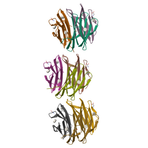
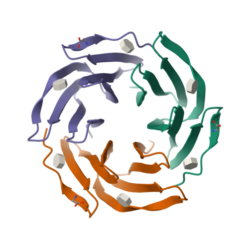
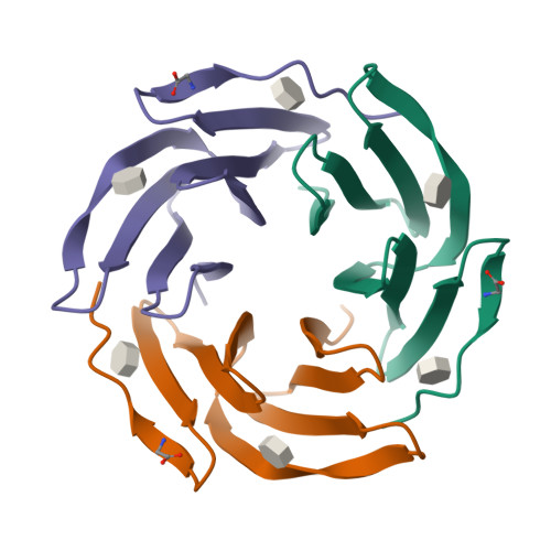
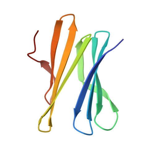
    6T99, 6T9A, 6T9B

  • PubMed Abstract: 

    The molecular recognition of carbohydrates by proteins plays a key role in many biological processes including immune response, pathogen entry into a cell, and cell-cell adhesion (e.g., in cancer metastasis). Carbohydrates interact with proteins mainly through hydrogen bonding, metal-ion-mediated interaction, and non-polar dispersion interactions. The role of dispersion-driven CH-π interactions (stacking) in protein-carbohydrate recognition has been underestimated for a long time considering the polar interactions to be the main forces for saccharide interactions. However, over the last few years it turns out that non-polar interactions are equally important. In this study, we analyzed the CH-π interactions employing bioinformatics (data mining, structural analysis), several experimental (isothermal titration calorimetry (ITC), X-ray crystallography), and computational techniques. The Protein Data Bank (PDB) has been used as a source of structural data. The PDB contains over 12 000 protein complexes with carbohydrates. Stacking interactions are very frequently present in such complexes (about 39 % of identified structures). The calculations and the ITC measurement results suggest that the CH-π stacking contribution to the overall binding energy ranges from 4 up to 8 kcal mol -1 . All the results show that the stacking CH-π interactions in protein-carbohydrate complexes can be considered to be a driving force of the binding in such complexes.


  • Organizational Affiliation

    Central European Institute of Technology, Masaryk University, Kamenice 5, 62500, Brno, Czech Republic.


Macromolecules
Find similar proteins by:  (by identity cutoff)  |  3D Structure
Entity ID: 1
MoleculeChains Sequence LengthOrganismDetailsImage
Fucose-binding lectin protein
A, B, C, D, E
90Ralstonia solanacearumMutation(s): 1 
Gene Names: RSP795_21825RSP799_05830RSP822_19650RUN39_v1_50103
UniProt
Find proteins for A0A0S4TLR1 (Ralstonia solanacearum)
Explore A0A0S4TLR1 
Go to UniProtKB:  A0A0S4TLR1
Entity Groups  
Sequence Clusters30% Identity50% Identity70% Identity90% Identity95% Identity100% Identity
UniProt GroupA0A0S4TLR1
Sequence Annotations
Expand
  • Reference Sequence
Small Molecules
Ligands 2 Unique
IDChains Name / Formula / InChI Key2D Diagram3D Interactions
MFU (Subject of Investigation/LOI)
Query on MFU

Download Ideal Coordinates CCD File 
AA [auth G]
BA [auth G]
DA [auth H]
EA [auth H]
GA [auth I]
methyl alpha-L-fucopyranoside
C7 H14 O5
OHWCAVRRXKJCRB-CXNFULCWSA-N
GLY
Query on GLY

Download Ideal Coordinates CCD File 
CA [auth G]
FA [auth H]
IA [auth I]
L [auth A]
O [auth B]
GLYCINE
C2 H5 N O2
DHMQDGOQFOQNFH-UHFFFAOYSA-N
Experimental Data & Validation

Experimental Data

  • Method: X-RAY DIFFRACTION
  • Resolution: 1.46 Å
  • R-Value Free:  0.211 (Depositor), 0.190 (DCC) 
  • R-Value Work:  0.184 (Depositor), 0.170 (DCC) 
  • R-Value Observed: 0.186 (Depositor) 
Space Group: P 1
Unit Cell:
Length ( Å )Angle ( ˚ )
a = 43.914α = 90.04
b = 44.048β = 91
c = 103.461γ = 119.67
Software Package:
Software NamePurpose
REFMACrefinement
SCALAdata scaling
PDB_EXTRACTdata extraction
XDSdata reduction
MOLREPphasing

Structure Validation

View Full Validation Report


Ligand Structure Quality Assessment 

Created with Raphaël 2.3.0Worse 01 BetterLigand structure goodness of fit to experimental dataBest fitted MFUClick on this verticalbar to view details

Entry History 

Deposition Data

Revision History  (Full details and data files)

  • Version 1.0: 2020-04-22
    Type: Initial release
  • Version 1.1: 2020-07-29
    Type: Remediation
    Reason: Carbohydrate remediation
    Changes: Data collection, Derived calculations, Structure summary
  • Version 1.2: 2020-08-12
    Changes: Database references, Structure summary
  • Version 1.3: 2020-12-16
    Changes: Database references
  • Version 1.4: 2024-01-24
    Changes: Data collection, Database references, Refinement description