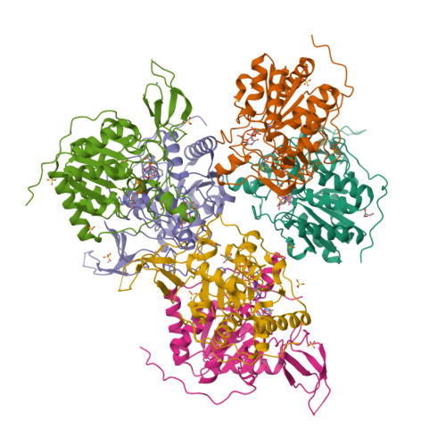
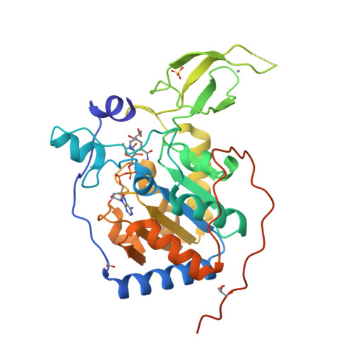
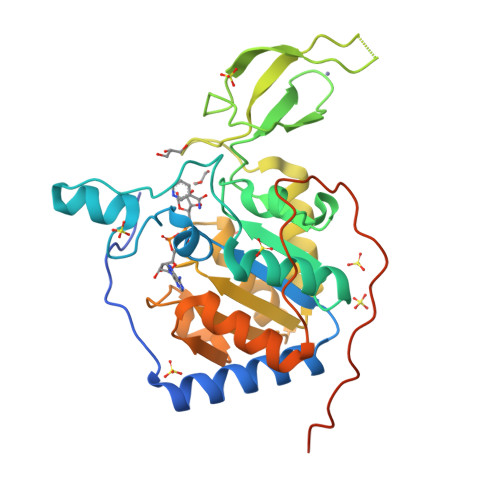
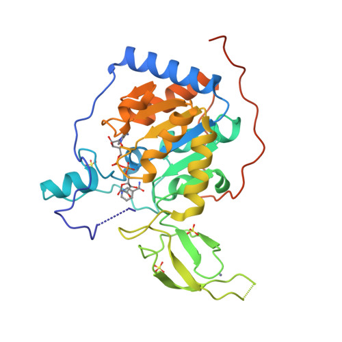
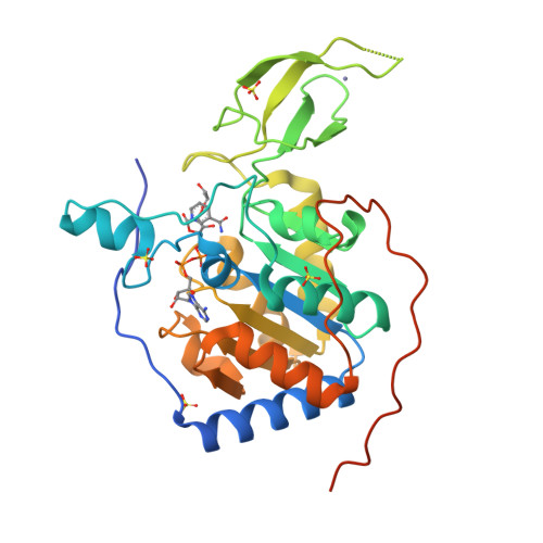
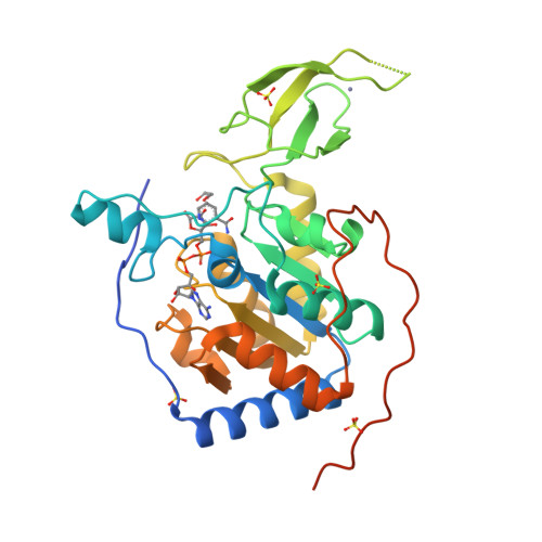
Binding site for activator MDL-801 on SIRT6.
You, W., Steegborn, C.(2021) Nat Chem Biol 17: 519-521
Experimental Data Snapshot
Starting Model: experimental
View more details
Entity ID: 1 | |||||
|---|---|---|---|---|---|
| Molecule | Chains | Sequence Length | Organism | Details | Image |
| NAD-dependent protein deacetylase sirtuin-6 | 316 | Homo sapiens | Mutation(s): 0 Gene Names: SIRT6, SIR2L6 EC: 2.3.1.286 (PDB Primary Data), 2.4.2 (UniProt), 2.3.1 (UniProt) |  | |
UniProt & NIH Common Fund Data Resources | |||||
Find proteins for Q8N6T7 (Homo sapiens) Explore Q8N6T7 Go to UniProtKB: Q8N6T7 | |||||
PHAROS: Q8N6T7 GTEx: ENSG00000077463 | |||||
Entity Groups | |||||
| Sequence Clusters | 30% Identity50% Identity70% Identity90% Identity95% Identity100% Identity | ||||
| UniProt Group | Q8N6T7 | ||||
Sequence AnnotationsExpand | |||||
| |||||
| Ligands 5 Unique | |||||
|---|---|---|---|---|---|
| ID | Chains | Name / Formula / InChI Key | 2D Diagram | 3D Interactions | |
| AR6 (Subject of Investigation/LOI) Query on AR6 | FA [auth D] G [auth A] N [auth B] NA [auth E] VA [auth F] | [(2R,3S,4R,5R)-5-(6-AMINOPURIN-9-YL)-3,4-DIHYDROXY-OXOLAN-2-YL]METHYL[HYDROXY-[[(2R,3S,4R,5S)-3,4,5-TRIHYDROXYOXOLAN-2-YL]METHOXY]PHOSPHORYL] HYDROGEN PHOSPHATE C15 H23 N5 O14 P2 SRNWOUGRCWSEMX-ZQSHOCFMSA-N |  | ||
| NCA Query on NCA | BA [auth C] HA [auth D] I [auth A] P [auth B] PA [auth E] | NICOTINAMIDE C6 H6 N2 O DFPAKSUCGFBDDF-UHFFFAOYSA-N |  | ||
| SO4 Query on SO4 | AB [auth F] BB [auth F] CA [auth C] DA [auth C] EA [auth C] | SULFATE ION O4 S QAOWNCQODCNURD-UHFFFAOYSA-L |  | ||
| GOL Query on GOL | IA [auth D] J [auth A] K [auth A] Q [auth B] QA [auth E] | GLYCEROL C3 H8 O3 PEDCQBHIVMGVHV-UHFFFAOYSA-N |  | ||
| ZN Query on ZN | AA [auth C] GA [auth D] H [auth A] O [auth B] OA [auth E] | ZINC ION Zn PTFCDOFLOPIGGS-UHFFFAOYSA-N |  | ||
| Length ( Å ) | Angle ( ˚ ) |
|---|---|
| a = 89.689 | α = 90 |
| b = 136.31 | β = 117.37 |
| c = 90.096 | γ = 90 |
| Software Name | Purpose |
|---|---|
| REFMAC | refinement |
| XDS | data reduction |
| XDS | data scaling |
| PHASER | phasing |
| PDB_EXTRACT | data extraction |
| Funding Organization | Location | Grant Number |
|---|---|---|
| German Research Foundation (DFG) | Germany | STE1701/15 |