6ZPC | pdb_00006zpc

Cyanophage S-2L HD phosphohydrolase (DatZ) bound to dATP


Experimental Data Snapshot

  • Method: X-RAY DIFFRACTION
  • Resolution: 1.27 Å
  • R-Value Free: 
    0.146 (Depositor), 0.150 (DCC) 
  • R-Value Work: 
    0.126 (Depositor), 0.130 (DCC) 
  • R-Value Observed: 
    0.127 (Depositor) 

wwPDB Validation   3D Report Full Report


Ligand Structure Quality Assessment 

Created with Raphaël 2.3.0Worse 01 BetterLigand structure goodness of fit to experimental dataBest fitted DTPClick on this verticalbar to view details

This is version 1.2 of the entry. See complete history


Literature

How cyanophage S-2L rejects adenine and incorporates 2-aminoadenine to saturate hydrogen bonding in its DNA.

Czernecki, D.Legrand, P.Tekpinar, M.Rosario, S.Kaminski, P.A.Delarue, M.

(2021) Nat Commun 12: 2420-2420

  • DOI: https://doi.org/10.1038/s41467-021-22626-x
  • Primary Citation of Related Structures:  
    6ZP9, 6ZPA, 6ZPB, 6ZPC

  • PubMed Abstract: 

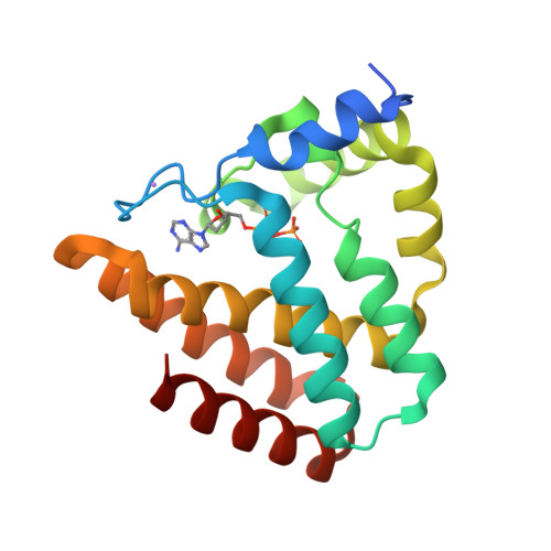
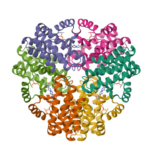
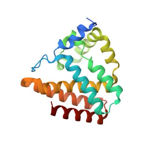
    Bacteriophages have long been known to use modified bases in their DNA to prevent cleavage by the host's restriction endonucleases. Among them, cyanophage S-2L is unique because its genome has all its adenines (A) systematically replaced by 2-aminoadenines (Z). Here, we identify a member of the PrimPol family as the sole possible polymerase of S-2L and we find it can incorporate both A and Z in front of a T. Its crystal structure at 1.5 Å resolution confirms that there is no structural element in the active site that could lead to the rejection of A in front of T. To resolve this contradiction, we show that a nearby gene is a triphosphohydolase specific of dATP (DatZ), that leaves intact all other dNTPs, including dZTP. This explains the absence of A in S-2L genome. Crystal structures of DatZ with various ligands, including one at sub-angstrom resolution, allow to describe its mechanism as a typical two-metal-ion mechanism and to set the stage for its engineering.


  • Organizational Affiliation

    Unit of Structural Dynamics of Biological Macromolecules, CNRS UMR 3528, 25-28 rue du Docteur Roux, Institut Pasteur, Paris, France.


Macromolecules
Find similar proteins by:  (by identity cutoff)  |  3D Structure
Entity ID: 1
MoleculeChains Sequence LengthOrganismDetailsImage
DatZ181Cyanophage S-2LMutation(s): 0 
UniProt
Find proteins for A0A8I3AZS8 (Cyanophage S-2L)
Explore A0A8I3AZS8 
Go to UniProtKB:  A0A8I3AZS8
Entity Groups  
Sequence Clusters30% Identity50% Identity70% Identity90% Identity95% Identity100% Identity
UniProt GroupA0A8I3AZS8
Sequence Annotations
Expand
  • Reference Sequence
Experimental Data & Validation

Experimental Data

  • Method: X-RAY DIFFRACTION
  • Resolution: 1.27 Å
  • R-Value Free:  0.146 (Depositor), 0.150 (DCC) 
  • R-Value Work:  0.126 (Depositor), 0.130 (DCC) 
  • R-Value Observed: 0.127 (Depositor) 
Space Group: H 3 2
Unit Cell:
Length ( Å )Angle ( ˚ )
a = 141.65α = 90
b = 141.65β = 90
c = 53.73γ = 120
Software Package:
Software NamePurpose
PHENIXrefinement
XDSdata reduction
XDSdata scaling
SHELXCDphasing

Structure Validation

View Full Validation Report


Ligand Structure Quality Assessment 

Created with Raphaël 2.3.0Worse 01 BetterLigand structure goodness of fit to experimental dataBest fitted DTPClick on this verticalbar to view details

Entry History 

Deposition Data

Revision History  (Full details and data files)

  • Version 1.0: 2021-03-03
    Type: Initial release
  • Version 1.1: 2021-05-05
    Changes: Database references
  • Version 1.2: 2024-05-15
    Changes: Data collection, Database references