7E7F | pdb_00007e7f

Human CYP11B1 mutant in complex with metyrapone


Experimental Data Snapshot

  • Method: X-RAY DIFFRACTION
  • Resolution: 1.40 Å
  • R-Value Free: 
    0.163 (Depositor), 0.160 (DCC) 
  • R-Value Work: 
    0.140 (Depositor), 0.140 (DCC) 
  • R-Value Observed: 
    0.141 (Depositor) 

Starting Model: experimental
View more details

wwPDB Validation   3D Report Full Report


Ligand Structure Quality Assessment 

Created with Raphaël 2.3.0Worse 01 BetterLigand structure goodness of fit to experimental dataBest fitted MYTClick on this verticalbar to view details

This is version 1.1 of the entry. See complete history


Literature

Spatially restricted substrate-binding site of cortisol-synthesizing CYP11B1 limits multiple hydroxylations and hinders aldosterone synthesis.

Mukai, K.Sugimoto, H.Kamiya, K.Suzuki, R.Matsuura, T.Hishiki, T.Shimada, H.Shiro, Y.Suematsu, M.Kagawa, N.

(2021) Curr Res Struct Biol 3: 192-205

  • DOI: https://doi.org/10.1016/j.crstbi.2021.08.001
  • Primary Citation of Related Structures:  
    7E7F

  • PubMed Abstract: 

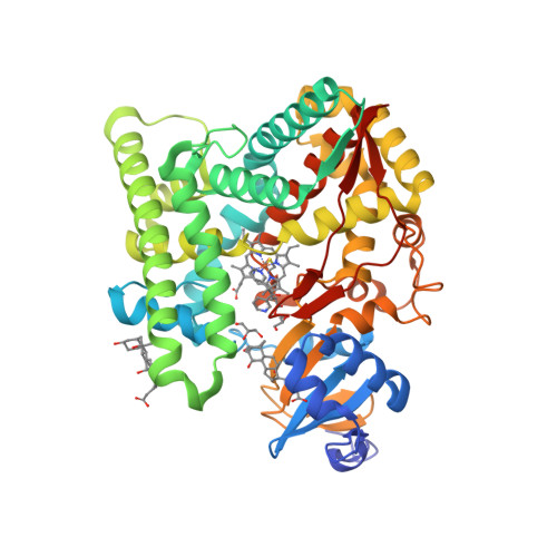
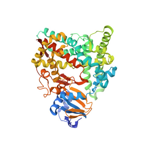
    Human cytochromes P450 11β (CYP11B1) and P450 aldo (CYP11B2) are monooxygenases that synthesize cortisol through steroid 11β-hydroxylation and aldosterone through a three-step process comprising 11β-hydroxylation and two 18-hydroxylations, respectively. CYP11B1 also catalyzes 18-monohydroxylation and 11β,18-dihydroxylation. To study the molecular basis of such catalytic divergence of the two enzymes, we examined a CYP11B1 mutant (Mt-CYP11B1) with amino acid replacements on the distal surface by determining the catalytic activities and crystal structure in the metyrapone-bound form at 1.4-Å resolution. Mt-CY11B1 retained both 11β-hydroxylase and 18-hydroxylase activities of the wild type (Wt-CYP11B1) but lacked 11β,18-dihydroxylase activity. Comparisons of the crystal structure of Mt-CYP11B1 to those of Wt-CYP11B1 and CYP11B2 that were already reported show that the mutation reduced the innermost space putatively surrounding the C3 side of substrate 11-deoxycorticosterone (DOC) bound to Wt-CYP11B1, while the corresponding space in CYP11B2 is enlarged markedly and accessible to bulk water through a channel. Molecular dynamics simulations of their DOC-bound forms supported the above findings and revealed that the enlarged space of CYP11B2 had a hydrogen bonding network involving water molecules that position DOC. Thus, upon positioning 11β-hydroxysteroid for 18-hydroxylation in their substrate-binding sites, steric hindrance could occur more strongly in Mt-CYP11B1 than in Wt-CYP11B1 but less in CYP11B2. Our investigation employing Mt-CYP11B1 sheds light on the divergence in structure and function between CYP11B1 and CYP11B2 and suggests that CYP11B1 with spatially-restricted substrate-binding site serves as 11β-hydroxylase, while CYP11B2 with spatially-extended substrate-binding site successively processes additional 18-hydroxylations to produce aldosterone.


  • Organizational Affiliation

    Department of Biochemistry, Keio University School of Medicine, Shinjuku, Tokyo 160-8582, Japan.


Macromolecules
Find similar proteins by:  (by identity cutoff)  |  3D Structure
Entity ID: 1
MoleculeChains Sequence LengthOrganismDetailsImage
Cytochrome P450 11B1, mitochondrial486Homo sapiensMutation(s): 6 
Gene Names: CYP11B1S11BH
EC: 1.14.15.4
UniProt & NIH Common Fund Data Resources
Find proteins for P15538 (Homo sapiens)
Explore P15538 
Go to UniProtKB:  P15538
PHAROS:  P15538
GTEx:  ENSG00000160882 
Entity Groups  
Sequence Clusters30% Identity50% Identity70% Identity90% Identity95% Identity100% Identity
UniProt GroupP15538
Sequence Annotations
Expand
  • Reference Sequence
Small Molecules
Ligands 4 Unique
IDChains Name / Formula / InChI Key2D Diagram3D Interactions
HEM
Query on HEM

Download Ideal Coordinates CCD File 
B [auth A]PROTOPORPHYRIN IX CONTAINING FE
C34 H32 Fe N4 O4
KABFMIBPWCXCRK-RGGAHWMASA-L
CHD
Query on CHD

Download Ideal Coordinates CCD File 
D [auth A],
E [auth A]
CHOLIC ACID
C24 H40 O5
BHQCQFFYRZLCQQ-OELDTZBJSA-N
MYT (Subject of Investigation/LOI)
Query on MYT

Download Ideal Coordinates CCD File 
C [auth A]METYRAPONE
C14 H14 N2 O
FJLBFSROUSIWMA-UHFFFAOYSA-N
GOL
Query on GOL

Download Ideal Coordinates CCD File 
F [auth A],
G [auth A]
GLYCEROL
C3 H8 O3
PEDCQBHIVMGVHV-UHFFFAOYSA-N
Binding Affinity Annotations 
IDSourceBinding Affinity
MYT BindingDB:  7E7F IC50: min: 5, max: 208 (nM) from 7 assay(s)
Experimental Data & Validation

Experimental Data

  • Method: X-RAY DIFFRACTION
  • Resolution: 1.40 Å
  • R-Value Free:  0.163 (Depositor), 0.160 (DCC) 
  • R-Value Work:  0.140 (Depositor), 0.140 (DCC) 
  • R-Value Observed: 0.141 (Depositor) 
Space Group: P 21 21 21
Unit Cell:
Length ( Å )Angle ( ˚ )
a = 75.18α = 90
b = 84.6β = 90
c = 85.61γ = 90
Software Package:
Software NamePurpose
XSCALEdata scaling
REFMACrefinement
PDB_EXTRACTdata extraction
ARP/wARPdata reduction
PHASERphasing

Structure Validation

View Full Validation Report


Ligand Structure Quality Assessment 

Created with Raphaël 2.3.0Worse 01 BetterLigand structure goodness of fit to experimental dataBest fitted MYTClick on this verticalbar to view details

Entry History & Funding Information

Deposition Data


Funding OrganizationLocationGrant Number
Japan Society for the Promotion of Science (JSPS)Japan26461387
Japan Society for the Promotion of Science (JSPS)Japan17K09890

Revision History  (Full details and data files)

  • Version 1.0: 2022-01-05
    Type: Initial release
  • Version 1.1: 2023-11-29
    Changes: Data collection, Refinement description