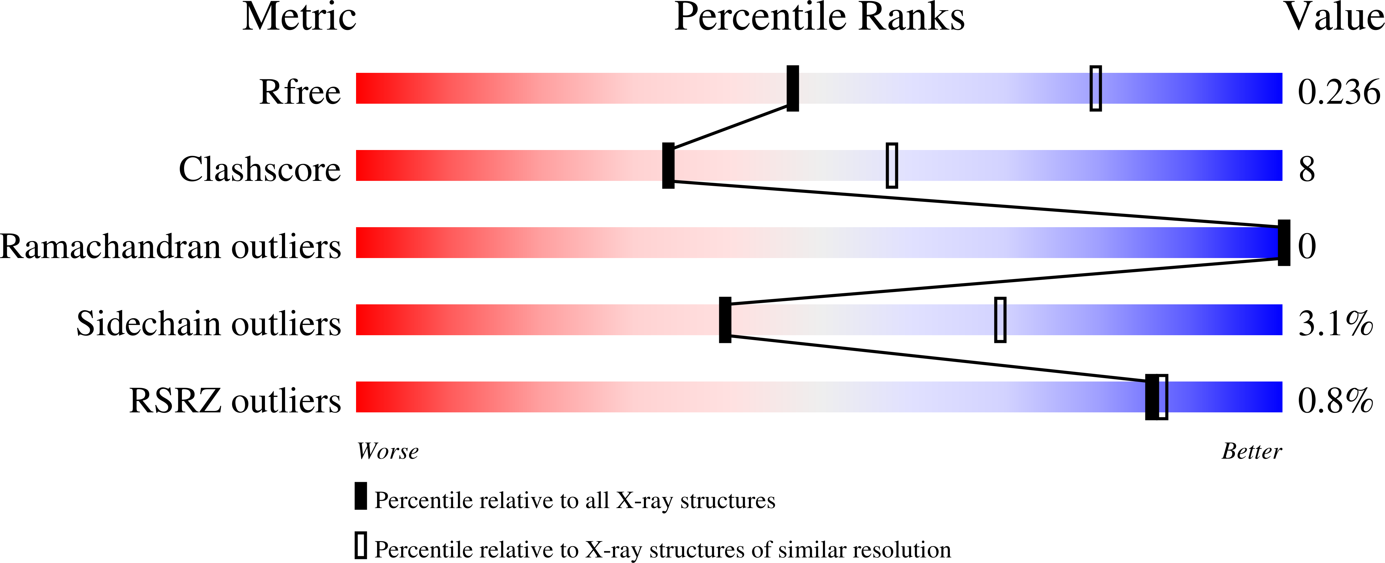
7KMA

Crystal structure of eif2Balpha with a ligand.


Experimental Data Snapshot

  • Method: X-RAY DIFFRACTION
  • Resolution: 2.70 Å
  • R-Value Free: 0.238 
  • R-Value Work: 0.191 
  • R-Value Observed: 0.193 

Starting Model: experimental
View more details

wwPDB Validation   3D Report Full Report


Ligand Structure Quality Assessment 


This is version 1.1 of the entry. See complete history


Literature

Sugar phosphate activation of the stress sensor eIF2B.

Hao, Q.Heo, J.M.Nocek, B.P.Hicks, K.G.Stoll, V.S.Remarcik, C.Hackett, S.LeBon, L.Jain, R.Eaton, D.Rutter, J.Wong, Y.L.Sidrauski, C.

(2021) Nat Commun 12: 3440-3440

  • DOI: https://doi.org/10.1038/s41467-021-23836-z
  • Primary Citation of Related Structures:  
    7KMA, 7KMF

  • PubMed Abstract: 

    The multi-subunit translation initiation factor eIF2B is a control node for protein synthesis. eIF2B activity is canonically modulated through stress-responsive phosphorylation of its substrate eIF2. The eIF2B regulatory subcomplex is evolutionarily related to sugar-metabolizing enzymes, but the biological relevance of this relationship was unknown. To identify natural ligands that might regulate eIF2B, we conduct unbiased binding- and activity-based screens followed by structural studies. We find that sugar phosphates occupy the ancestral catalytic site in the eIF2Bα subunit, promote eIF2B holoenzyme formation and enhance enzymatic activity towards eIF2. A mutant in the eIF2Bα ligand pocket that causes Vanishing White Matter disease fails to engage and is not stimulated by sugar phosphates. These data underscore the importance of allosteric metabolite modulation for proper eIF2B function. We propose that eIF2B evolved to couple nutrient status via sugar phosphate sensing with the rate of protein synthesis, one of the most energetically costly cellular processes.


  • Organizational Affiliation

    Calico Life Sciences LLC, South San Francisco, CA, USA.


Macromolecules
Find similar proteins by:  (by identity cutoff)  |  3D Structure
Entity ID: 1
MoleculeChains Sequence LengthOrganismDetailsImage
Translation initiation factor eIF-2B subunit alpha
A, B, C, D, E
A, B, C, D, E, F, G, H
313Homo sapiensMutation(s): 0 
Gene Names: EIF2B1EIF2BA
UniProt & NIH Common Fund Data Resources
Find proteins for Q14232 (Homo sapiens)
Explore Q14232 
Go to UniProtKB:  Q14232
PHAROS:  Q14232
GTEx:  ENSG00000111361 
Entity Groups  
Sequence Clusters30% Identity50% Identity70% Identity90% Identity95% Identity100% Identity
UniProt GroupQ14232
Sequence Annotations
Expand
  • Reference Sequence
Small Molecules
Ligands 2 Unique
IDChains Name / Formula / InChI Key2D Diagram3D Interactions
M6P (Subject of Investigation/LOI)
Query on M6P

Download Ideal Coordinates CCD File 
I [auth A]
K [auth B]
L [auth C]
M [auth D]
N [auth E]
I [auth A],
K [auth B],
L [auth C],
M [auth D],
N [auth E],
O [auth F],
P [auth G],
Q [auth H]
6-O-phosphono-alpha-D-mannopyranose
C6 H13 O9 P
NBSCHQHZLSJFNQ-PQMKYFCFSA-N
ACT (Subject of Investigation/LOI)
Query on ACT

Download Ideal Coordinates CCD File 
J [auth A]ACETATE ION
C2 H3 O2
QTBSBXVTEAMEQO-UHFFFAOYSA-M
Experimental Data & Validation

Experimental Data

  • Method: X-RAY DIFFRACTION
  • Resolution: 2.70 Å
  • R-Value Free: 0.238 
  • R-Value Work: 0.191 
  • R-Value Observed: 0.193 
  • Space Group: P 1 21 1
Unit Cell:
Length ( Å )Angle ( ˚ )
a = 71.2α = 90
b = 155.54β = 103.893
c = 140.11γ = 90
Software Package:
Software NamePurpose
PHENIXrefinement
XDSdata reduction
pointlessdata scaling
MOLREPphasing

Structure Validation

View Full Validation Report


Ligand Structure Quality Assessment 


Entry History 

Deposition Data

Revision History  (Full details and data files)

  • Version 1.0: 2021-07-21
    Type: Initial release
  • Version 1.1: 2023-10-18
    Changes: Advisory, Data collection, Database references, Refinement description