7NOU

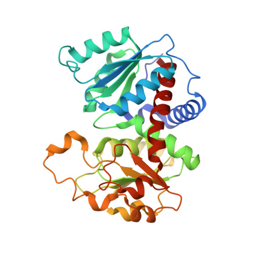
Crystal structure of Mycobacterium tuberculosis ArgF in complex with (3,5-dichlorophenyl)boronic acid.


Experimental Data Snapshot

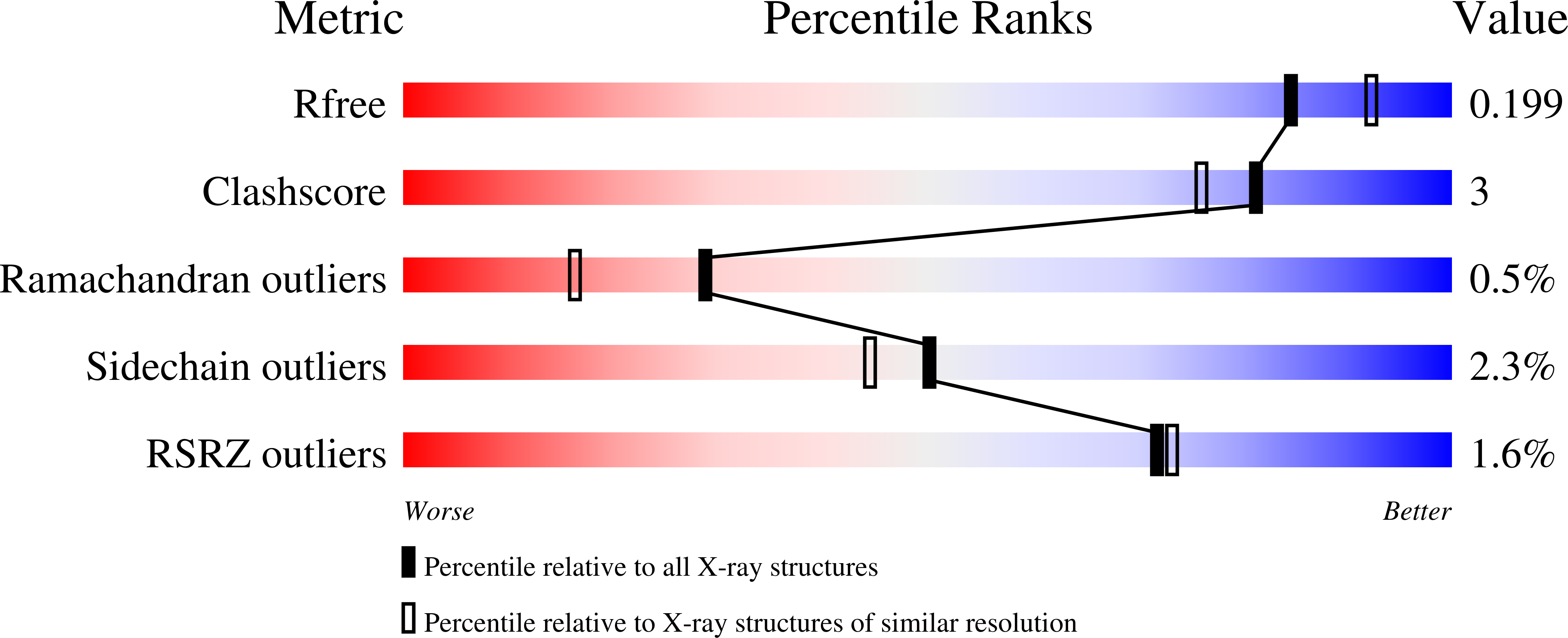
  • Method: X-RAY DIFFRACTION
  • Resolution: 1.98 Å
  • R-Value Free: 0.201 
  • R-Value Work: 0.174 
  • R-Value Observed: 0.176 

Starting Model: experimental
View more details

wwPDB Validation   3D Report Full Report


Ligand Structure Quality Assessment 


This is version 1.1 of the entry. See complete history


Literature

A fragment-based approach to assess the ligandability of ArgB, ArgC, ArgD and ArgF in the L-arginine biosynthetic pathway of Mycobacterium tuberculosis

Gupta, P.Thomas, S.E.Zaidan, S.A.Pasillas, M.A.Cory-Wright, J.Sebastian-Perez, V.Burgess, A.Cattermole, E.Meghir, C.Abell, C.Coyne, A.G.Jacobs, W.R.Blundell, T.L.Tiwari, S.Mendes, V.

(2021) Comput Struct Biotechnol J 19: 3491-3506


Macromolecules
Find similar proteins by:  (by identity cutoff)  |  3D Structure
Entity ID: 1
MoleculeChains Sequence LengthOrganismDetailsImage
Ornithine carbamoyltransferase
A, B, C, D, E
A, B, C, D, E, F
309Mycobacterium tuberculosis H37RvMutation(s): 0 
Gene Names: argFRv1656MTCY06H11.21
EC: 2.1.3.3
UniProt
Find proteins for P9WIT9 (Mycobacterium tuberculosis (strain ATCC 25618 / H37Rv))
Explore P9WIT9 
Go to UniProtKB:  P9WIT9
Entity Groups  
Sequence Clusters30% Identity50% Identity70% Identity90% Identity95% Identity100% Identity
UniProt GroupP9WIT9
Sequence Annotations
Expand
  • Reference Sequence
Small Molecules
Ligands 2 Unique
IDChains Name / Formula / InChI Key2D Diagram3D Interactions
NMR (Subject of Investigation/LOI)
Query on NMR

Download Ideal Coordinates CCD File 
G [auth A]
I [auth A]
J [auth A]
K [auth A]
M [auth C]
G [auth A],
I [auth A],
J [auth A],
K [auth A],
M [auth C],
N [auth C],
O [auth D],
P [auth D],
Q [auth D],
T [auth F],
U [auth F]
[3,5-bis(chloranyl)phenyl]-oxidanyl-oxidanylidene-boron
C6 H4 B Cl2 O2
QFYDXKOJHIFUIC-UHFFFAOYSA-N
PO4
Query on PO4

Download Ideal Coordinates CCD File 
H [auth A],
L [auth B],
R [auth E],
S [auth E]
PHOSPHATE ION
O4 P
NBIIXXVUZAFLBC-UHFFFAOYSA-K
Experimental Data & Validation

Experimental Data

  • Method: X-RAY DIFFRACTION
  • Resolution: 1.98 Å
  • R-Value Free: 0.201 
  • R-Value Work: 0.174 
  • R-Value Observed: 0.176 
  • Space Group: P 1 21 1
Unit Cell:
Length ( Å )Angle ( ˚ )
a = 92.299α = 90
b = 144.662β = 117.8
c = 97.756γ = 90
Software Package:
Software NamePurpose
PHENIXrefinement
Aimlessdata scaling
PHASERphasing
PDB_EXTRACTdata extraction
XDSdata reduction

Structure Validation

View Full Validation Report


Ligand Structure Quality Assessment 


Entry History & Funding Information

Deposition Data


Funding OrganizationLocationGrant Number
Bill & Melinda Gates FoundationUnited StatesOPP1158806

Revision History  (Full details and data files)

  • Version 1.0: 2021-06-30
    Type: Initial release
  • Version 1.1: 2024-01-31
    Changes: Data collection, Database references, Refinement description