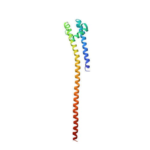
7PGI

NaVAb1p (bicelles)

  • Classification: MEMBRANE PROTEIN
  • Organism(s): Alcanivorax borkumensis SK2
  • Expression System: Escherichia coli
  • Mutation(s): No 
  • Membrane Protein: Yes  OPMPDBTM

  • Deposited: 2021-08-14 Released: 2022-06-08 
  • Deposition Author(s): Lolicato, M., Arrigoni, C.
  • Funding Organization(s): National Institutes of Health/National Heart, Lung, and Blood Institute (NIH/NHLBI), National Institutes of Health/National Institute on Deafness and Other Communication Disorders (NIH/NIDCD), National Institutes of Health/National Institute of General Medical Sciences (NIH/NIGMS)

Experimental Data Snapshot

  • Method: X-RAY DIFFRACTION
  • Resolution: 3.64 Å
  • R-Value Free: 0.303 
  • R-Value Work: 0.287 
  • R-Value Observed: 0.288 

Starting Model: experimental
View more details

wwPDB Validation   3D Report Full Report


This is version 1.3 of the entry. See complete history


Literature

Quaternary structure independent folding of voltage-gated ion channel pore domain subunits.

Arrigoni, C.Lolicato, M.Shaya, D.Rohaim, A.Findeisen, F.Fong, L.K.Colleran, C.M.Dominik, P.Kim, S.S.Schuermann, J.P.DeGrado, W.F.Grabe, M.Kossiakoff, A.A.Minor Jr., D.L.

(2022) Nat Struct Mol Biol 29: 537-548

  • DOI: https://doi.org/10.1038/s41594-022-00775-x
  • Primary Citation of Related Structures:  
    7PG8, 7PGB, 7PGF, 7PGG, 7PGH, 7PGI

  • PubMed Abstract: 

    Every voltage-gated ion channel (VGIC) has a pore domain (PD) made from four subunits, each comprising an antiparallel transmembrane helix pair bridged by a loop. The extent to which PD subunit structure requires quaternary interactions is unclear. Here, we present crystal structures of a set of bacterial voltage-gated sodium channel (BacNa V ) 'pore only' proteins that reveal a surprising collection of non-canonical quaternary arrangements in which the PD tertiary structure is maintained. This context-independent structural robustness, supported by molecular dynamics simulations, indicates that VGIC-PD tertiary structure is independent of quaternary interactions. This fold occurs throughout the VGIC superfamily and in diverse transmembrane and soluble proteins. Strikingly, characterization of PD subunit-binding Fabs indicates that non-canonical quaternary PD conformations can occur in full-length VGICs. Together, our data demonstrate that the VGIC-PD is an autonomously folded unit. This property has implications for VGIC biogenesis, understanding functional states, de novo channel design, and VGIC structural origins.


  • Organizational Affiliation

    Cardiovascular Research Institute, University of California, San Francisco, CA, USA.


Macromolecules
Find similar proteins by:  (by identity cutoff)  |  3D Structure
Entity ID: 1
MoleculeChains Sequence LengthOrganismDetailsImage
Ion transport protein
A, B, C, D, E
A, B, C, D, E, F, G, H
150Alcanivorax borkumensis SK2Mutation(s): 0 
Gene Names: ABO_1668
Membrane Entity: Yes 
UniProt
Find proteins for Q0VNY2 (Alcanivorax borkumensis (strain ATCC 700651 / DSM 11573 / NCIMB 13689 / SK2))
Explore Q0VNY2 
Go to UniProtKB:  Q0VNY2
Entity Groups  
Sequence Clusters30% Identity50% Identity70% Identity90% Identity95% Identity100% Identity
UniProt GroupQ0VNY2
Sequence Annotations
Expand
  • Reference Sequence
Small Molecules
Ligands 3 Unique
IDChains Name / Formula / InChI Key2D Diagram3D Interactions
ACT
Query on ACT

Download Ideal Coordinates CCD File 
I [auth A]
L [auth C]
M [auth D]
O [auth E]
R [auth G]
I [auth A],
L [auth C],
M [auth D],
O [auth E],
R [auth G],
S [auth H]
ACETATE ION
C2 H3 O2
QTBSBXVTEAMEQO-UHFFFAOYSA-M
MG
Query on MG

Download Ideal Coordinates CCD File 
N [auth E]MAGNESIUM ION
Mg
JLVVSXFLKOJNIY-UHFFFAOYSA-N
NA
Query on NA

Download Ideal Coordinates CCD File 
J [auth A],
K [auth A],
P [auth F],
Q [auth G]
SODIUM ION
Na
FKNQFGJONOIPTF-UHFFFAOYSA-N
Experimental Data & Validation

Experimental Data

  • Method: X-RAY DIFFRACTION
  • Resolution: 3.64 Å
  • R-Value Free: 0.303 
  • R-Value Work: 0.287 
  • R-Value Observed: 0.288 
  • Space Group: I 21 21 21
Unit Cell:
Length ( Å )Angle ( ˚ )
a = 178.18α = 90
b = 191.8β = 90
c = 192.32γ = 90
Software Package:
Software NamePurpose
PHENIXrefinement
PDB_EXTRACTdata extraction
MOSFLMdata reduction
SCALAdata scaling
Cootmodel building
PHASERphasing

Structure Validation

View Full Validation Report


Entry History & Funding Information

Deposition Data


Funding OrganizationLocationGrant Number
National Institutes of Health/National Heart, Lung, and Blood Institute (NIH/NHLBI)United States--
National Institutes of Health/National Institute on Deafness and Other Communication Disorders (NIH/NIDCD)United States--
National Institutes of Health/National Institute of General Medical Sciences (NIH/NIGMS)United States--

Revision History  (Full details and data files)

  • Version 1.0: 2022-06-08
    Type: Initial release
  • Version 1.1: 2022-06-15
    Changes: Database references
  • Version 1.2: 2022-06-29
    Changes: Database references
  • Version 1.3: 2024-01-31
    Changes: Data collection, Refinement description