7QBS

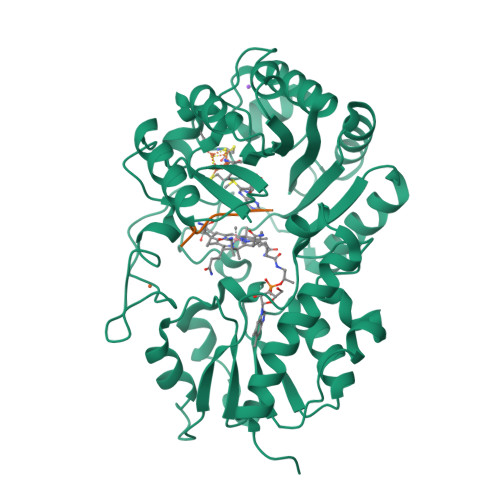
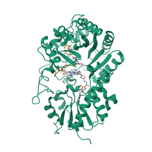
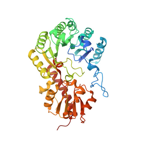
B12-dependent radical SAM methyltransferase, Mmp10 with [4Fe-4S] cluster, cobalamin, S-adenosyl-L-methionine, and peptide bound.


Experimental Data Snapshot

  • Method: X-RAY DIFFRACTION
  • Resolution: 2.33 Å
  • R-Value Free: 0.235 
  • R-Value Work: 0.182 
  • R-Value Observed: 0.185 

wwPDB Validation   3D Report Full Report


Ligand Structure Quality Assessment 


This is version 1.3 of the entry. See complete history


Literature

Crystallographic snapshots of a B 12 -dependent radical SAM methyltransferase.

Fyfe, C.D.Bernardo-Garcia, N.Fradale, L.Grimaldi, S.Guillot, A.Brewee, C.Chavas, L.M.G.Legrand, P.Benjdia, A.Berteau, O.

(2022) Nature 602: 336-342

  • DOI: https://doi.org/10.1038/s41586-021-04355-9
  • Primary Citation of Related Structures:  
    7QBS, 7QBT, 7QBU, 7QBV

  • PubMed Abstract: 

    By catalysing the microbial formation of methane, methyl-coenzyme M reductase has a central role in the global levels of this greenhouse gas 1,2 . The activity of methyl-coenzyme M reductase is profoundly affected by several unique post-translational modifications 3-6 , such as  a unique C-methylation reaction catalysed by methanogenesis marker protein 10 (Mmp10), a radical S-adenosyl-L-methionine (SAM) enzyme 7,8 . Here we report the spectroscopic investigation and atomic resolution structure of Mmp10 from Methanosarcina acetivorans, a unique B 12 (cobalamin)-dependent radical SAM enzyme 9 . The structure of Mmp10 reveals a unique enzyme architecture with four metallic centres and critical structural features involved in the control of catalysis. In addition, the structure of the enzyme-substrate complex offers a glimpse into a B 12 -dependent radical SAM enzyme in a precatalytic state. By combining electron paramagnetic resonance spectroscopy, structural biology and biochemistry, our study illuminates the mechanism by which the emerging superfamily of B 12 -dependent radical SAM enzymes catalyse chemically challenging alkylation reactions and identifies distinctive active site rearrangements to provide a structural rationale for the dual use of the SAM cofactor for radical and nucleophilic chemistry.


  • Organizational Affiliation

    Université Paris-Saclay, INRAE, AgroParisTech, Micalis Institute, ChemSyBio, Jouy-en-Josas, France.


Macromolecules
Find similar proteins by:  (by identity cutoff)  |  3D Structure
Entity ID: 1
MoleculeChains Sequence LengthOrganismDetailsImage
Methyl coenzyme M reductase-arginine methyltransferase Mmp10436Methanosarcina acetivoransMutation(s): 0 
Gene Names: mmp10HA338_00275
EC: 2.1.1 (PDB Primary Data), 2.1.1.379 (UniProt)
UniProt
Find proteins for Q8THG6 (Methanosarcina acetivorans (strain ATCC 35395 / DSM 2834 / JCM 12185 / C2A))
Explore Q8THG6 
Go to UniProtKB:  Q8THG6
Entity Groups  
Sequence Clusters30% Identity50% Identity70% Identity90% Identity95% Identity100% Identity
UniProt GroupQ8THG6
Sequence Annotations
Expand
  • Reference Sequence

Find similar proteins by:  Sequence   |   3D Structure  

Entity ID: 2
MoleculeChains Sequence LengthOrganismDetailsImage
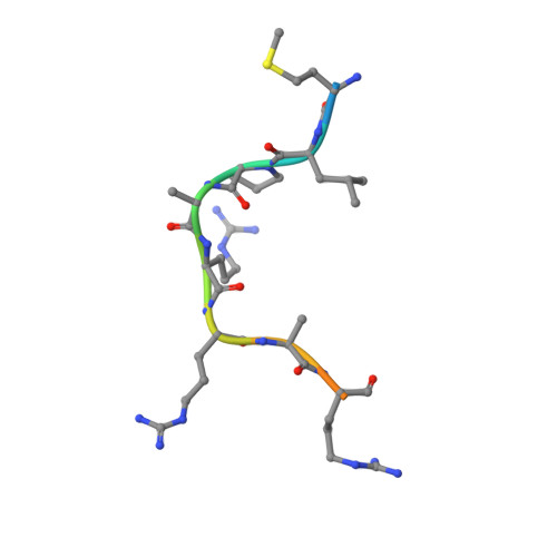
Peptide from Methyl-coenzyme M reductase subunit alpha from Methanosarcina acetivorans13Methanosarcina acetivoransMutation(s): 0 
EC: 2.8.4.1
UniProt
Find proteins for Q8THH1 (Methanosarcina acetivorans (strain ATCC 35395 / DSM 2834 / JCM 12185 / C2A))
Explore Q8THH1 
Go to UniProtKB:  Q8THH1
Entity Groups  
Sequence Clusters30% Identity50% Identity70% Identity90% Identity95% Identity100% Identity
UniProt GroupQ8THH1
Sequence Annotations
Expand
  • Reference Sequence
Small Molecules
Ligands 5 Unique
IDChains Name / Formula / InChI Key2D Diagram3D Interactions
COB (Subject of Investigation/LOI)
Query on COB

Download Ideal Coordinates CCD File 
F [auth A]CO-METHYLCOBALAMIN
C63 H91 Co N13 O14 P
BMOGZGJBQIUQJG-BYLSPCLQSA-N
SAM (Subject of Investigation/LOI)
Query on SAM

Download Ideal Coordinates CCD File 
E [auth A]S-ADENOSYLMETHIONINE
C15 H22 N6 O5 S
MEFKEPWMEQBLKI-FCKMPRQPSA-N
SF4 (Subject of Investigation/LOI)
Query on SF4

Download Ideal Coordinates CCD File 
C [auth A]IRON/SULFUR CLUSTER
Fe4 S4
LJBDFODJNLIPKO-UHFFFAOYSA-N
FE (Subject of Investigation/LOI)
Query on FE

Download Ideal Coordinates CCD File 
D [auth A]FE (III) ION
Fe
VTLYFUHAOXGGBS-UHFFFAOYSA-N
NA (Subject of Investigation/LOI)
Query on NA

Download Ideal Coordinates CCD File 
G [auth A]SODIUM ION
Na
FKNQFGJONOIPTF-UHFFFAOYSA-N
Experimental Data & Validation

Experimental Data

  • Method: X-RAY DIFFRACTION
  • Resolution: 2.33 Å
  • R-Value Free: 0.235 
  • R-Value Work: 0.182 
  • R-Value Observed: 0.185 
  • Space Group: P 63
Unit Cell:
Length ( Å )Angle ( ˚ )
a = 119.29α = 90
b = 119.29β = 90
c = 68.56γ = 120
Software Package:
Software NamePurpose
BUSTERrefinement
XDSdata reduction
XSCALEdata scaling
SHELXCDphasing
SHARPphasing
SOLOMONphasing

Structure Validation

View Full Validation Report


Ligand Structure Quality Assessment 


Entry History & Funding Information

Deposition Data


Funding OrganizationLocationGrant Number
European Research Council (ERC)France617053
Agence Nationale de la Recherche (ANR)FranceANR-17-CE11-0014

Revision History  (Full details and data files)

  • Version 1.0: 2022-02-02
    Type: Initial release
  • Version 1.1: 2022-02-16
    Changes: Database references
  • Version 1.2: 2022-02-23
    Changes: Database references
  • Version 1.3: 2024-06-19
    Changes: Data collection