7VSO

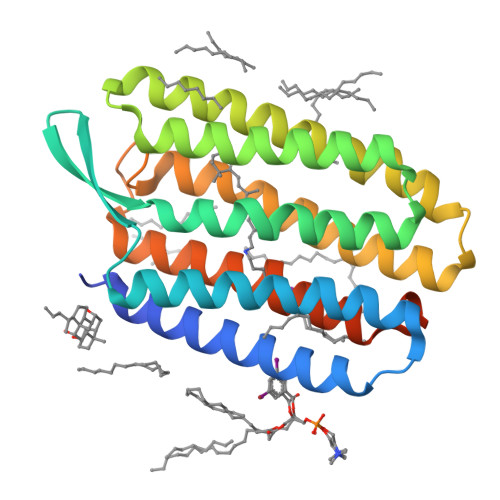
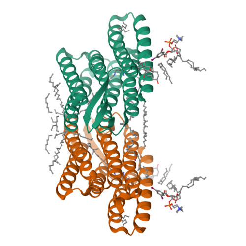
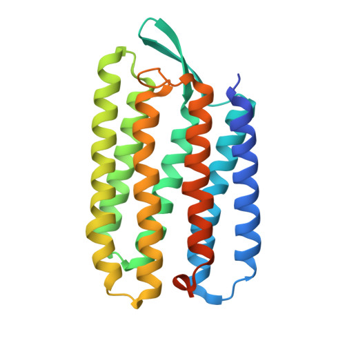
Serial Femtosecond Crystallography (SFX) of Ground State Bacteriorhodopsin Crystallized from Bicelles in Complex with HAD16 Determined Using 7-keV X-ray Free Electron Laser (XFEL) at SACLA


Experimental Data Snapshot

  • Method: X-RAY DIFFRACTION
  • Resolution: 2.35 Å
  • R-Value Free: 0.210 
  • R-Value Work: 0.158 
  • R-Value Observed: 0.160 

Starting Model: experimental
View more details

wwPDB Validation   3D Report Full Report


Ligand Structure Quality Assessment 

Created with Raphaël 2.3.0Worse 01 BetterLigand structure goodness of fit to experimental dataBest fitted 7YHClick on this verticalbar to view detailsBest fitted CPSClick on this verticalbar to view detailsBest fitted RETClick on this verticalbar to view detailsBest fitted R16Click on this verticalbar to view detailsBest fitted D12Click on this verticalbar to view detailsBest fitted D10Click on this verticalbar to view detailsBest fitted DD9Click on this verticalbar to view detailsBest fitted OCTClick on this verticalbar to view detailsBest fitted HP6Click on this verticalbar to view details

This is version 1.2 of the entry. See complete history


Literature

Heavy Atom Detergent/Lipid Combined X-ray Crystallography for Elucidating the Structure-Function Relationships of Membrane Proteins.

Hanashima, S.Nakane, T.Mizohata, E.

(2021) Membranes (Basel) 11

  • DOI: https://doi.org/10.3390/membranes11110823
  • Primary Citation of Related Structures:  
    7VSO

  • PubMed Abstract: 

    Membrane proteins reside in the lipid bilayer of biomembranes and the structure and function of these proteins are closely related to their interactions with lipid molecules. Structural analyses of interactions between membrane proteins and lipids or detergents that constitute biological or artificial model membranes are important for understanding the functions and physicochemical properties of membrane proteins and biomembranes. Determination of membrane protein structures is much more difficult when compared with that of soluble proteins, but the development of various new technologies has accelerated the elucidation of the structure-function relationship of membrane proteins. This review summarizes the development of heavy atom derivative detergents and lipids that can be used for structural analysis of membrane proteins and their interactions with detergents/lipids, including their application with X-ray free-electron laser crystallography.


  • Organizational Affiliation

    Department of Chemistry, Graduate School of Science, Osaka University, 1-1 Machikaneyama, Toyonaka 563-0043, Japan.


Macromolecules
Find similar proteins by:  (by identity cutoff)  |  3D Structure
Entity ID: 1
MoleculeChains Sequence LengthOrganismDetailsImage
Bacteriorhodopsin249Halobacterium salinarumMutation(s): 0 
Membrane Entity: Yes 
UniProt
Find proteins for P02945 (Halobacterium salinarum (strain ATCC 700922 / JCM 11081 / NRC-1))
Explore P02945 
Go to UniProtKB:  P02945
Entity Groups  
Sequence Clusters30% Identity50% Identity70% Identity90% Identity95% Identity100% Identity
UniProt GroupP02945
Sequence Annotations
Expand
  • Reference Sequence
Small Molecules
Ligands 10 Unique
IDChains Name / Formula / InChI Key2D Diagram3D Interactions
7YH (Subject of Investigation/LOI)
Query on 7YH

Download Ideal Coordinates CCD File 
C [auth A]2-[[(2R)-2-(3-bromanyl-5-iodanyl-phenyl)carbonyloxy-3-tetradecanoyloxy-propoxy]-oxidanyl-phosphoryl]oxyethyl-trimethyl-azanium
C29 H49 Br I N O8 P
RDKBFTQTYYTUKD-HHHXNRCGSA-O
CPS (Subject of Investigation/LOI)
Query on CPS

Download Ideal Coordinates CCD File 
E [auth A]3-[(3-CHOLAMIDOPROPYL)DIMETHYLAMMONIO]-1-PROPANESULFONATE
C32 H58 N2 O7 S
UMCMPZBLKLEWAF-BCTGSCMUSA-N
RET (Subject of Investigation/LOI)
Query on RET

Download Ideal Coordinates CCD File 
B [auth A]RETINAL
C20 H28 O
NCYCYZXNIZJOKI-OVSJKPMPSA-N
R16 (Subject of Investigation/LOI)
Query on R16

Download Ideal Coordinates CCD File 
I [auth A],
K [auth A],
S [auth A]
HEXADECANE
C16 H34
DCAYPVUWAIABOU-UHFFFAOYSA-N
D12 (Subject of Investigation/LOI)
Query on D12

Download Ideal Coordinates CCD File 
H [auth A],
Q [auth A],
R [auth A]
DODECANE
C12 H26
SNRUBQQJIBEYMU-UHFFFAOYSA-N
D10 (Subject of Investigation/LOI)
Query on D10

Download Ideal Coordinates CCD File 
J [auth A],
M [auth A]
DECANE
C10 H22
DIOQZVSQGTUSAI-UHFFFAOYSA-N
DD9 (Subject of Investigation/LOI)
Query on DD9

Download Ideal Coordinates CCD File 
P [auth A]nonane
C9 H20
BKIMMITUMNQMOS-UHFFFAOYSA-N
IOD (Subject of Investigation/LOI)
Query on IOD

Download Ideal Coordinates CCD File 
D [auth A]IODIDE ION
I
XMBWDFGMSWQBCA-UHFFFAOYSA-M
OCT (Subject of Investigation/LOI)
Query on OCT

Download Ideal Coordinates CCD File 
F [auth A],
U [auth A]
N-OCTANE
C8 H18
TVMXDCGIABBOFY-UHFFFAOYSA-N
HP6 (Subject of Investigation/LOI)
Query on HP6

Download Ideal Coordinates CCD File 
G [auth A],
L [auth A],
N [auth A],
O [auth A],
T [auth A]
HEPTANE
C7 H16
IMNFDUFMRHMDMM-UHFFFAOYSA-N
Experimental Data & Validation

Experimental Data

Unit Cell:
Length ( Å )Angle ( ˚ )
a = 46.2α = 90
b = 103β = 90
c = 128.7γ = 90
Software Package:
Software NamePurpose
REFMACrefinement
CrystFELdata reduction
CrystFELdata scaling
Cootmodel building

Structure Validation

View Full Validation Report


Ligand Structure Quality Assessment 

Created with Raphaël 2.3.0Worse 01 BetterLigand structure goodness of fit to experimental dataBest fitted 7YHClick on this verticalbar to view detailsBest fitted CPSClick on this verticalbar to view detailsBest fitted RETClick on this verticalbar to view detailsBest fitted R16Click on this verticalbar to view detailsBest fitted D12Click on this verticalbar to view detailsBest fitted D10Click on this verticalbar to view detailsBest fitted DD9Click on this verticalbar to view detailsBest fitted OCTClick on this verticalbar to view detailsBest fitted HP6Click on this verticalbar to view details

Entry History & Funding Information

Deposition Data


Funding OrganizationLocationGrant Number
Japan Science and TechnologyJapanJPMJPR17GB

Revision History  (Full details and data files)

  • Version 1.0: 2022-02-16
    Type: Initial release
  • Version 1.1: 2023-09-06
    Changes: Data collection, Database references
  • Version 1.2: 2023-11-29
    Changes: Refinement description