7Y4A | pdb_00007y4a

Crystal structure of human ELMO1 RBD-RhoG complex


Experimental Data Snapshot

  • Method: X-RAY DIFFRACTION
  • Resolution: 1.60 Å
  • R-Value Free: 
    0.235 (Depositor), 0.240 (DCC) 
  • R-Value Work: 
    0.198 (Depositor), 0.200 (DCC) 
  • R-Value Observed: 
    0.198 (Depositor) 

Starting Models: experimental
View more details

wwPDB Validation   3D Report Full Report


Ligand Structure Quality Assessment 

Created with Raphaël 2.3.0Worse 01 BetterLigand structure goodness of fit to experimental dataBest fitted GDPClick on this verticalbar to view detailsBest fitted PO4Click on this verticalbar to view details

This is version 1.2 of the entry. See complete history


Literature

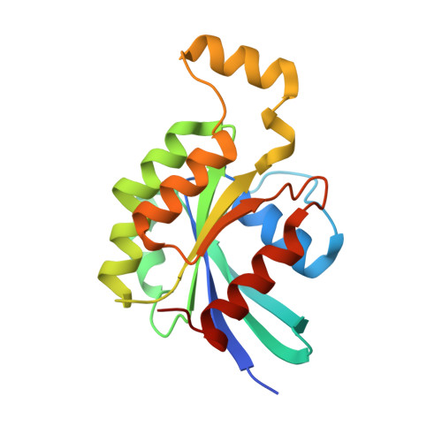
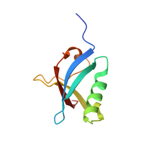
Targeting Ras-binding domain of ELMO1 by computational nanobody design.

Tam, C.Kukimoto-Niino, M.Miyata-Yabuki, Y.Tsuda, K.Mishima-Tsumagari, C.Ihara, K.Inoue, M.Yonemochi, M.Hanada, K.Matsumoto, T.Shirouzu, M.Zhang, K.Y.J.

(2023) Commun Biol 6: 284-284

  • DOI: https://doi.org/10.1038/s42003-023-04657-w
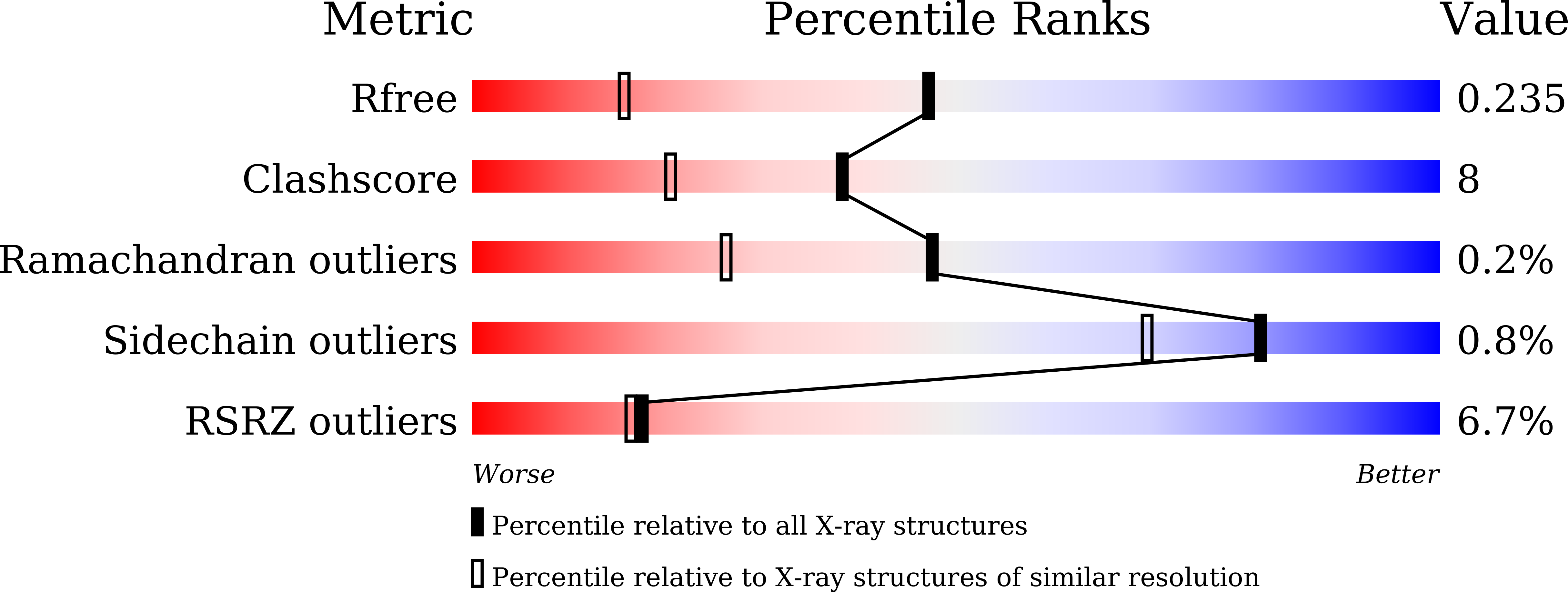
  • Primary Citation of Related Structures:  
    6JPP, 7Y4A

  • PubMed Abstract: 

    The control of cell movement through manipulation of cytoskeletal structure has therapeutic prospects notably in the development of novel anti-metastatic drugs. In this study, we determine the structure of Ras-binding domain (RBD) of ELMO1, a protein involved in cytoskeletal regulation, both alone and in complex with the activator RhoG and verify its targetability through computational nanobody design. Using our dock-and-design approach optimized with native-like initial pose selection, we obtain Nb01, a detectable binder from scratch in the first-round design. An affinity maturation step guided by structure-activity relationship at the interface generates 23 Nb01 sequence variants and 17 of them show enhanced binding to ELMO1-RBD and are modeled to form major spatial overlaps with RhoG. The best binder, Nb29, inhibited ELMO1-RBD/RhoG interaction. Molecular dynamics simulation of the flexibility of CDR2 and CDR3 of Nb29 reveal the design of stabilizing mutations at the CDR-framework junctions potentially confers the affinity enhancement.


  • Organizational Affiliation

    Laboratory for Structural Bioinformatics, Center for Biosystems Dynamics Research, RIKEN, 1-7-22 Suehiro, Tsurumi, Yokohama, Kanagawa, 230-0045, Japan.


Macromolecules
Find similar proteins by:  (by identity cutoff)  |  3D Structure
Entity ID: 1
MoleculeChains Sequence LengthOrganismDetailsImage
Rho-related GTP-binding protein RhoG
A, C, E, G
191Homo sapiensMutation(s): 0 
Gene Names: RHOGARHG
UniProt & NIH Common Fund Data Resources
Find proteins for P84095 (Homo sapiens)
Explore P84095 
Go to UniProtKB:  P84095
PHAROS:  P84095
GTEx:  ENSG00000177105 
Entity Groups  
Sequence Clusters30% Identity50% Identity70% Identity90% Identity95% Identity100% Identity
UniProt GroupP84095
Sequence Annotations
Expand
  • Reference Sequence
Find similar proteins by:  (by identity cutoff)  |  3D Structure
Entity ID: 2
MoleculeChains Sequence LengthOrganismDetailsImage
Engulfment and cell motility protein 1
B, D, F, H
83Homo sapiensMutation(s): 0 
Gene Names: ELMO1
UniProt & NIH Common Fund Data Resources
Find proteins for Q92556 (Homo sapiens)
Explore Q92556 
Go to UniProtKB:  Q92556
PHAROS:  Q92556
GTEx:  ENSG00000155849 
Entity Groups  
Sequence Clusters30% Identity50% Identity70% Identity90% Identity95% Identity100% Identity
UniProt GroupQ92556
Sequence Annotations
Expand
  • Reference Sequence
Small Molecules
Ligands 3 Unique
IDChains Name / Formula / InChI Key2D Diagram3D Interactions
GDP (Subject of Investigation/LOI)
Query on GDP

Download Ideal Coordinates CCD File 
K [auth A],
N [auth C],
Q [auth E],
T [auth G]
GUANOSINE-5'-DIPHOSPHATE
C10 H15 N5 O11 P2
QGWNDRXFNXRZMB-UUOKFMHZSA-N
PO4 (Subject of Investigation/LOI)
Query on PO4

Download Ideal Coordinates CCD File 
J [auth A],
M [auth C],
P [auth E],
S [auth G]
PHOSPHATE ION
O4 P
NBIIXXVUZAFLBC-UHFFFAOYSA-K
MG (Subject of Investigation/LOI)
Query on MG

Download Ideal Coordinates CCD File 
I [auth A],
L [auth C],
O [auth E],
R [auth G]
MAGNESIUM ION
Mg
JLVVSXFLKOJNIY-UHFFFAOYSA-N
Experimental Data & Validation

Experimental Data

  • Method: X-RAY DIFFRACTION
  • Resolution: 1.60 Å
  • R-Value Free:  0.235 (Depositor), 0.240 (DCC) 
  • R-Value Work:  0.198 (Depositor), 0.200 (DCC) 
  • R-Value Observed: 0.198 (Depositor) 
Space Group: P 1 21 1
Unit Cell:
Length ( Å )Angle ( ˚ )
a = 74.72α = 90
b = 63.75β = 100.5
c = 115.33γ = 90
Software Package:
Software NamePurpose
PHENIXrefinement
XDSdata reduction
XDSdata scaling
PHASERphasing

Structure Validation

View Full Validation Report


Ligand Structure Quality Assessment 

Created with Raphaël 2.3.0Worse 01 BetterLigand structure goodness of fit to experimental dataBest fitted GDPClick on this verticalbar to view detailsBest fitted PO4Click on this verticalbar to view details

Entry History & Funding Information

Deposition Data


Funding OrganizationLocationGrant Number
Japan Society for the Promotion of Science (JSPS)Japan--

Revision History  (Full details and data files)

  • Version 1.0: 2023-03-15
    Type: Initial release
  • Version 1.1: 2023-03-29
    Changes: Database references
  • Version 1.2: 2023-11-29
    Changes: Data collection, Refinement description