


Structure of the mouse 8-oxoguanine DNA Glycosylase mOGG1 in complex with ligand TH013545
Scaletti, E.R., Stenmark, P.To be published.
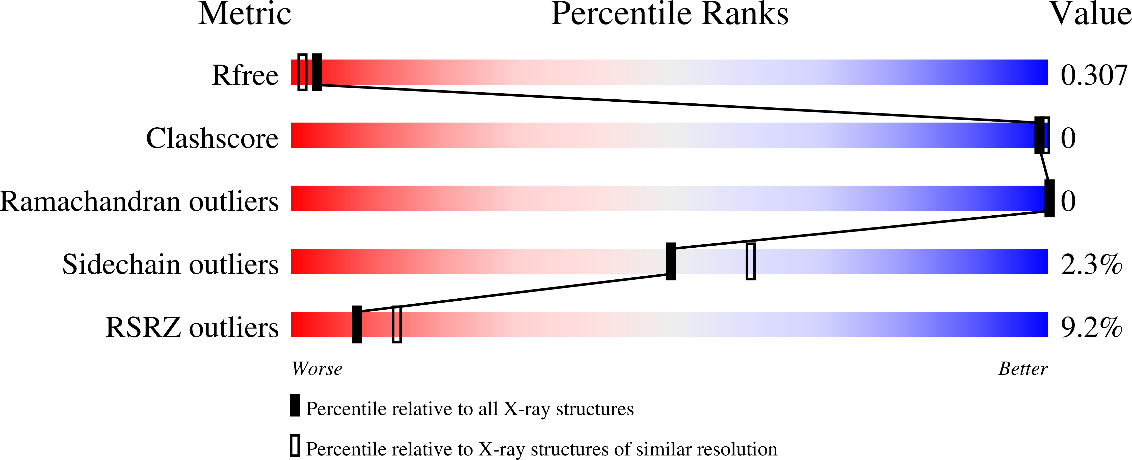
Experimental Data Snapshot
Starting Model: experimental
View more details
Entity ID: 1 | |||||
|---|---|---|---|---|---|
| Molecule | Chains | Sequence Length | Organism | Details | Image |
| N-glycosylase/DNA lyase | A [auth AAA], B [auth BBB], C [auth CCC] | 318 | Mus musculus | Mutation(s): 0 Gene Names: Ogg1 EC: 3.2.2 (PDB Primary Data), 4.2.99.18 (PDB Primary Data) | 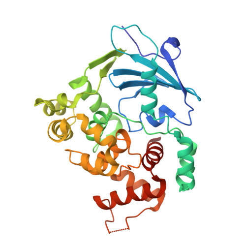 |
UniProt | |||||
Find proteins for O08760 (Mus musculus) Explore O08760 Go to UniProtKB: O08760 | |||||
Entity Groups | |||||
| Sequence Clusters | 30% Identity50% Identity70% Identity90% Identity95% Identity100% Identity | ||||
| UniProt Group | O08760 | ||||
Sequence AnnotationsExpand | |||||
| |||||
| Ligands 3 Unique | |||||
|---|---|---|---|---|---|
| ID | Chains | Name / Formula / InChI Key | 2D Diagram | 3D Interactions | |
| I9U (Subject of Investigation/LOI) Query on I9U | D [auth AAA] | 2-[4-(3,5-dimethylpyrazol-1-yl)-2,6-bis(fluoranyl)phenyl]-~{N}-(4,5,6,7-tetrahydro-1,2-benzoxazol-3-yl)ethanamide C20 H20 F2 N4 O2 KHMWCMCBIAGTAK-UHFFFAOYSA-N |  | ||
| GOL Query on GOL | E [auth AAA], G [auth BBB] | GLYCEROL C3 H8 O3 PEDCQBHIVMGVHV-UHFFFAOYSA-N |  | ||
| NI Query on NI | F [auth AAA] | NICKEL (II) ION Ni VEQPNABPJHWNSG-UHFFFAOYSA-N |  | ||
| Length ( Å ) | Angle ( ˚ ) |
|---|---|
| a = 80.995 | α = 90 |
| b = 81.831 | β = 90 |
| c = 168.952 | γ = 90 |
| Software Name | Purpose |
|---|---|
| REFMAC | refinement |
| DIALS | data reduction |
| Aimless | data scaling |
| PHASER | phasing |
| Funding Organization | Location | Grant Number |
|---|---|---|
| Cancerfonden | Sweden | -- |
RCSB PDB (citation) is hosted by
RCSB PDB is a member of the

