7QBU

B12-dependent radical SAM methyltransferase, Mmp10 with [4Fe-4S] cluster, cobalamin, and S-methyl-5'-thioadenosine bound.


Experimental Data Snapshot

  • Method: X-RAY DIFFRACTION
  • Resolution: 2.30 Å
  • R-Value Free: 
    0.225 (Depositor), 0.210 (DCC) 
  • R-Value Work: 
    0.191 (Depositor), 0.180 (DCC) 
  • R-Value Observed: 
    0.193 (Depositor) 

Starting Model: experimental
View more details

wwPDB Validation   3D Report Full Report


Ligand Structure Quality Assessment 

Created with Raphaël 2.3.0Worse 01 BetterLigand structure goodness of fit to experimental dataBest fitted COBClick on this verticalbar to view detailsBest fitted MTAClick on this verticalbar to view details

This is version 1.3 of the entry. See complete history


Literature

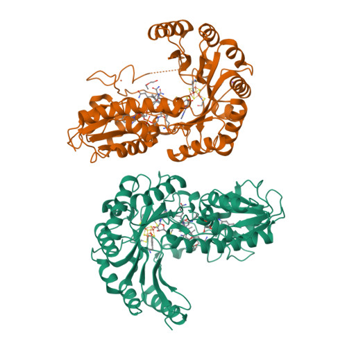
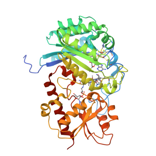
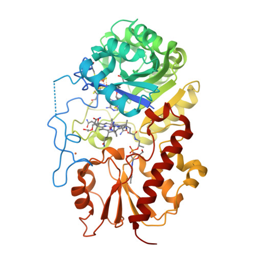
Crystallographic snapshots of a B 12 -dependent radical SAM methyltransferase.

Fyfe, C.D.Bernardo-Garcia, N.Fradale, L.Grimaldi, S.Guillot, A.Brewee, C.Chavas, L.M.G.Legrand, P.Benjdia, A.Berteau, O.

(2022) Nature 602: 336-342

  • DOI: https://doi.org/10.1038/s41586-021-04355-9
  • Primary Citation of Related Structures:  
    7QBS, 7QBT, 7QBU, 7QBV

  • PubMed Abstract: 

    By catalysing the microbial formation of methane, methyl-coenzyme M reductase has a central role in the global levels of this greenhouse gas 1,2 . The activity of methyl-coenzyme M reductase is profoundly affected by several unique post-translational modifications 3-6 , such as  a unique C-methylation reaction catalysed by methanogenesis marker protein 10 (Mmp10), a radical S-adenosyl-L-methionine (SAM) enzyme 7,8 . Here we report the spectroscopic investigation and atomic resolution structure of Mmp10 from Methanosarcina acetivorans, a unique B 12 (cobalamin)-dependent radical SAM enzyme 9 . The structure of Mmp10 reveals a unique enzyme architecture with four metallic centres and critical structural features involved in the control of catalysis. In addition, the structure of the enzyme-substrate complex offers a glimpse into a B 12 -dependent radical SAM enzyme in a precatalytic state. By combining electron paramagnetic resonance spectroscopy, structural biology and biochemistry, our study illuminates the mechanism by which the emerging superfamily of B 12 -dependent radical SAM enzymes catalyse chemically challenging alkylation reactions and identifies distinctive active site rearrangements to provide a structural rationale for the dual use of the SAM cofactor for radical and nucleophilic chemistry.


  • Organizational Affiliation

    Université Paris-Saclay, INRAE, AgroParisTech, Micalis Institute, ChemSyBio, Jouy-en-Josas, France.


Macromolecules
Find similar proteins by:  (by identity cutoff)  |  3D Structure
Entity ID: 1
MoleculeChains Sequence LengthOrganismDetailsImage
Methyl coenzyme M reductase-arginine methyltransferase Mmp10
A, B
436Methanosarcina acetivoransMutation(s): 0 
Gene Names: mmp10HA338_00275
EC: 2.1.1 (PDB Primary Data), 2.1.1.379 (UniProt)
UniProt
Find proteins for Q8THG6 (Methanosarcina acetivorans (strain ATCC 35395 / DSM 2834 / JCM 12185 / C2A))
Explore Q8THG6 
Go to UniProtKB:  Q8THG6
Entity Groups  
Sequence Clusters30% Identity50% Identity70% Identity90% Identity95% Identity100% Identity
UniProt GroupQ8THG6
Sequence Annotations
Expand
  • Reference Sequence
Small Molecules
Ligands 6 Unique
IDChains Name / Formula / InChI Key2D Diagram3D Interactions
COB (Subject of Investigation/LOI)
Query on COB

Download Ideal Coordinates CCD File 
F [auth A],
L [auth B]
CO-METHYLCOBALAMIN
C63 H91 Co N13 O14 P
BMOGZGJBQIUQJG-BYLSPCLQSA-N
SF4 (Subject of Investigation/LOI)
Query on SF4

Download Ideal Coordinates CCD File 
C [auth A],
I [auth B]
IRON/SULFUR CLUSTER
Fe4 S4
LJBDFODJNLIPKO-UHFFFAOYSA-N
MTA (Subject of Investigation/LOI)
Query on MTA

Download Ideal Coordinates CCD File 
E [auth A],
K [auth B]
5'-DEOXY-5'-METHYLTHIOADENOSINE
C11 H15 N5 O3 S
WUUGFSXJNOTRMR-IOSLPCCCSA-N
PEG
Query on PEG

Download Ideal Coordinates CCD File 
H [auth A],
N [auth B]
DI(HYDROXYETHYL)ETHER
C4 H10 O3
MTHSVFCYNBDYFN-UHFFFAOYSA-N
FE (Subject of Investigation/LOI)
Query on FE

Download Ideal Coordinates CCD File 
D [auth A],
J [auth B]
FE (III) ION
Fe
VTLYFUHAOXGGBS-UHFFFAOYSA-N
NA (Subject of Investigation/LOI)
Query on NA

Download Ideal Coordinates CCD File 
G [auth A],
M [auth B]
SODIUM ION
Na
FKNQFGJONOIPTF-UHFFFAOYSA-N
Experimental Data & Validation

Experimental Data

  • Method: X-RAY DIFFRACTION
  • Resolution: 2.30 Å
  • R-Value Free:  0.225 (Depositor), 0.210 (DCC) 
  • R-Value Work:  0.191 (Depositor), 0.180 (DCC) 
  • R-Value Observed: 0.193 (Depositor) 
Space Group: P 21 21 21
Unit Cell:
Length ( Å )Angle ( ˚ )
a = 77.779α = 90
b = 79.493β = 90
c = 151.659γ = 90
Software Package:
Software NamePurpose
BUSTERrefinement
XDSdata reduction
XSCALEdata scaling
REFMACphasing

Structure Validation

View Full Validation Report


Ligand Structure Quality Assessment 

Created with Raphaël 2.3.0Worse 01 BetterLigand structure goodness of fit to experimental dataBest fitted COBClick on this verticalbar to view detailsBest fitted MTAClick on this verticalbar to view details

Entry History & Funding Information

Deposition Data


Funding OrganizationLocationGrant Number
European Research Council (ERC)France617053
Agence Nationale de la Recherche (ANR)FranceANR-17-CE11-0014

Revision History  (Full details and data files)

  • Version 1.0: 2022-02-02
    Type: Initial release
  • Version 1.1: 2022-02-16
    Changes: Database references
  • Version 1.2: 2022-02-23
    Changes: Database references
  • Version 1.3: 2024-01-31
    Changes: Data collection, Refinement description