8BAC

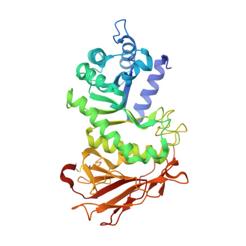
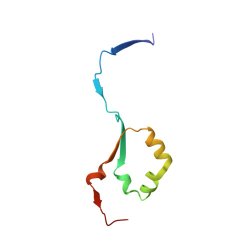
Crystal structure of human heparanase in complex with competitive inhibitor GD05


Experimental Data Snapshot

  • Method: X-RAY DIFFRACTION
  • Resolution: 2.05 Å
  • R-Value Free: 0.267 
  • R-Value Work: 0.224 

Starting Model: experimental
View more details

wwPDB Validation   3D Report Full Report


Ligand Structure Quality Assessment 


This is version 1.2 of the entry. See complete history


Literature

Synthesis of Uronic Acid 1-Azasugars as Putative Inhibitors of alpha-Iduronidase, beta-Glucuronidase and Heparanase.

Doherty, G.G.Ler, G.J.M.Wimmer, N.Bernhardt, P.V.Ashmus, R.A.Vocadlo, D.J.Armstrong, Z.Davies, G.J.Maccarana, M.Li, J.P.Kayal, Y.Ferro, V.

(2023) Chembiochem 24: e202200619-e202200619

  • DOI: https://doi.org/10.1002/cbic.202200619
  • Primary Citation of Related Structures:  
    8BAC

  • PubMed Abstract: 

    1-Azasugar analogues of l-iduronic acid (l-IdoA) and d-glucuronic acid (d-GlcA) and their corresponding enantiomers have been synthesized as potential pharmacological chaperones for mucopolysaccharidosis I (MPS I), a lysosomal storage disease caused by mutations in the gene encoding α-iduronidase (IDUA). The compounds were efficiently synthesized in nine or ten steps from d- or l-arabinose, and the structures were confirmed by X-ray crystallographic analysis of key intermediates. All compounds were inactive against IDUA, although l-IdoA-configured 8 moderately inhibited β-glucuronidase (β-GLU). The d-GlcA-configured 9 was a potent inhibitor of β-GLU and a moderate inhibitor of the endo-β-glucuronidase heparanase. Co-crystallization of 9 with heparanase revealed that the endocyclic nitrogen of 9 forms close interactions with both the catalytic acid and catalytic nucleophile.


  • Organizational Affiliation

    School of Chemistry & Molecular Biosciences, The University of Queensland, Brisbane, Queensland, 4072, Australia.


Macromolecules
Find similar proteins by:  (by identity cutoff)  |  3D Structure
Entity ID: 1
MoleculeChains Sequence LengthOrganismDetailsImage
Heparanase 50 kDa subunitA [auth AAA]389Homo sapiensMutation(s): 1 
Gene Names: HPSEHEPHPAHPA1HPR1HPSE1HSE1
EC: 3.2.1.166
UniProt & NIH Common Fund Data Resources
Find proteins for Q9Y251 (Homo sapiens)
Explore Q9Y251 
Go to UniProtKB:  Q9Y251
PHAROS:  Q9Y251
GTEx:  ENSG00000173083 
Entity Groups  
Sequence Clusters30% Identity50% Identity70% Identity90% Identity95% Identity100% Identity
UniProt GroupQ9Y251
Glycosylation
Glycosylation Sites: 2Go to GlyGen: Q9Y251-1
Sequence Annotations
Expand
  • Reference Sequence
Find similar proteins by:  (by identity cutoff)  |  3D Structure
Entity ID: 2
MoleculeChains Sequence LengthOrganismDetailsImage
Heparanase 8 kDa subunitB [auth BBB]74Homo sapiensMutation(s): 0 
Gene Names: HPSEHEPHPAHPA1HPR1HPSE1HSE1
EC: 3.2.1.166
UniProt & NIH Common Fund Data Resources
Find proteins for Q9Y251 (Homo sapiens)
Explore Q9Y251 
Go to UniProtKB:  Q9Y251
PHAROS:  Q9Y251
GTEx:  ENSG00000173083 
Entity Groups  
Sequence Clusters30% Identity50% Identity70% Identity90% Identity95% Identity100% Identity
UniProt GroupQ9Y251
Sequence Annotations
Expand
  • Reference Sequence
Experimental Data & Validation

Experimental Data

  • Method: X-RAY DIFFRACTION
  • Resolution: 2.05 Å
  • R-Value Free: 0.267 
  • R-Value Work: 0.224 
  • Space Group: P 1 21 1
Unit Cell:
Length ( Å )Angle ( ˚ )
a = 44.838α = 90
b = 71.188β = 98.375
c = 78.628γ = 90
Software Package:
Software NamePurpose
REFMACrefinement
xia2data reduction
xia2data scaling
PHASERphasing

Structure Validation

View Full Validation Report


Ligand Structure Quality Assessment 


Entry History & Funding Information

Deposition Data


Funding OrganizationLocationGrant Number
Biotechnology and Biological Sciences Research Council (BBSRC)United KingdomKen murray professorship

Revision History  (Full details and data files)

  • Version 1.0: 2023-03-01
    Type: Initial release
  • Version 1.1: 2024-02-07
    Changes: Data collection, Refinement description
  • Version 1.2: 2024-10-16
    Changes: Structure summary